
Pain in your pelvic region can be frustrating. Sometimes, the cause of pelvic pain isn’t immediately clear to your healthcare provider. This is why your provider recommended diagnostic imaging.
Whether your healthcare provider ordered an ultrasound or a CT scan, you should know that both diagnostic scans are excellent for finding the source of your pelvic pain.
But it might not be so easy for you to figure out which scan is better for your particular needs and your individual situation. We’ll take a closer look at ultrasounds and CT scans for pelvic pain, to help you understand why your provider has requested imaging, and give you the information you need to make an educated decision about your healthcare.
If I have pelvic pain, why did my healthcare provider recommend diagnostic imaging?
Pelvic pain could be caused by a variety of issues, including ovarian cysts, endometriosis, urinary tract infections (UTIs), digestive problems, and others.
It’s crucial to figure out the source to be able to address it effectively.
That’s where diagnostic imaging comes into play. Imaging techniques like ultrasounds and CT scans provide a non-invasive window into your body, allowing healthcare providers to identify any physical abnormalities that may be causing your discomfort.
What is an ultrasound?
An ultrasound is a form of imaging that uses sound waves to create images of soft tissues inside your body.
It’s similar to taking screenshots of what’s going on inside you, giving your healthcare provider real-time images.
An ultrasound of the pelvis can help your provider visualize your bladder, uterus (for women), prostate (for men), and other parts of your reproductive and urinary systems.
It’s safe, non-invasive, and doesn’t use radiation, which makes it a popular test for many people’s diagnostic imaging.

What is a CT scan?
A CT scan, or Computed Tomography scan, uses a combination of special equipment, advanced x-ray images and computer algorithms to provide more detailed, cross-sectional views of your body.
This kind of scan gives a comprehensive, 3D image of your pelvic area, allowing for a thorough examination of your organs and tissues.
CT scans can show both bone and soft tissues, including organs, muscles, and tumors, if present.
How are ultrasounds and CT scans different?
An ultrasound uses sound waves, and it’s ideal for real-time imaging, like assessing blood flow or fetal development in pregnant women. It’s also generally less expensive and doesn’t use any radiation.
CT scans, on the other hand, create a series of images that the machine takes from different angles. A CT can make highly detailed images, and can better visualize organs, blood vessels, and bones in the pelvic area. However, a CT scan does involve exposure to a small amount of radiation.
Do healthcare providers usually recommend an ultrasound or a CT for pelvic pain?
When it comes to diagnosing pelvic pain, both ultrasound and CT scans are commonly used, and each has its strengths.
The choice largely depends on your specific symptoms, overall health, and the suspected underlying condition.
An ultrasound is often the first-line diagnostic tool for pelvic pain, as it’s safe, readily available, and excellent at imaging soft tissues and fluid-filled structures, like cysts.
Your provider might recommend a CT scan next if the cause of your pelvic pain is still unclear after an ultrasound, or if they need a more detailed view of your body.
Remember, your healthcare provider will consider all of your healthcare info before recommending either of these diagnostic tools.
Understanding pelvic ultrasounds
During an ultrasound scan of the pelvis, an ultrasound probe is gently moved across your lower abdomen.
The sound waves that the probe produces will bounce back after hitting the internal organs, creating patterns from the echoes.
A computer then turns these echo patterns into images, allowing your healthcare provider to visualize your organs and structures in the pelvic region, in real time.
These ultrasound images can reveal abnormalities such as cysts, fibroids, or other potential causes of pelvic pain.
What are the advantages of using ultrasound for pelvic pain?
Ultrasound imaging is a non-invasive and painless procedure, which often makes it the first choice for initial investigation of pelvic pain.
Also, ultrasounds provide real-time imagery, which can be particularly beneficial in situations that require immediate observation, like checking blood flow in the pelvic organs.
Ultrasound imaging doesn’t use any radiation, making it a safe option for all patients, including those who might be pregnant.
It’s also widely available, and an ultrasound tends to be more affordable than a CT scan.
Another advantage is that ultrasounds are excellent at imaging soft tissues and fluid-filled structures, like ovarian cysts in women or prostate problems in men, which can be common causes of pelvic pain.
Are there any limitations or risks with using ultrasound for pelvic pain?
While ultrasound imaging is generally very safe and well-tolerated, it does have a few limitations.
One of the main limitations is that it may not provide the detailed images necessary to identify smaller abnormalities or complications within the pelvic organs.
Also, its effectiveness can sometimes be reduced in patients who have larger bodies or more abdominal fat, which could interfere with the scan.
Unlike other imaging methods, ultrasound may not capture certain areas as clearly, such as the deep pelvic organs or structures hidden behind the bowel.
Understanding CT scans for pelvic pain
A CT scan, short for Computed Tomography, is a sophisticated imaging technique that provides a more detailed and comprehensive view of the inside of your body compared to a traditional ultrasound.
The CT scan works by capturing multiple image slices of your body from different angles, which a computer then compiles into a detailed, cross-sectional 3D image.
For diagnosing pelvic pain, the CT scan gives a thorough examination of your pelvic area, including the reproductive system, urinary system, and lower digestive tract.
What are the advantages of using a CT scan for pelvic pain?
CT scans provide a higher resolution image than ultrasounds so they can detect smaller and more subtle abnormalities.
CT scans are excellent for identifying issues like kidney stones, diverticulitis, or tumors that could be causing pelvic pain.
Because of its cross-sectional view, a CT scan can provide a better look at the layers of the pelvic organs, which can be important if your healthcare provider is looking for issues such as complex ovarian cysts, abscesses, or other deep-seated pelvic disorders.
Usually, CT scans aren’t affected by the patient’s body size, unlike ultrasounds, which can sometimes struggle to produce clear images in larger people.
Are there any limitations or risks with using a CT scan for pelvic pain?
While CT scans are incredibly helpful, they do come with some limitations and potential risks.
CT scans expose you to a small amount of radiation, though it’s typically at a safe level, and the benefits often outweigh the risks. Check with your provider for more info.
Usually, pregnant women are advised to avoid CT scans to avoid any potential risk to the unborn baby.
People with kidney disease or certain allergies may not be suitable candidates for CT scans. That’s because CT scans often require a contrast material, which is a special dye that can improve the clarity of the images, and might cause reactions or complications in some people.
Also, CT scans can be more expensive than ultrasounds, and might not be the first-line diagnostic tool due to cost considerations.
Comparing ultrasound and CT for pelvic pain
Both ultrasounds and CT scans are comfortable and convenient procedures.
For an ultrasound, you’ll lie down while a healthcare professional gently moves a probe across your lower abdomen. There’s typically no pain involved, and the procedure itself can take anywhere from 15 minutes to an hour.
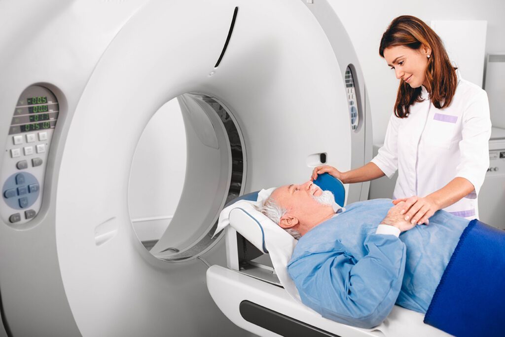
A CT scan involves lying down on a narrow table that slides into a large, doughnut-shaped machine.
While some people might feel claustrophobic during a CT, the scan is painless, and you’ll be in constant communication with a radiology technologist.
Which scan has quicker results, ultrasound or CT?
Both ultrasound and CT scans can produce images fairly quickly.
To get the best results, a radiology technologist usually needs to review these images before a diagnosis can be made, which might take a few days.
As such, the speed of getting results is more dependent on your healthcare provider’s office or facility procedures than on the imaging technique itself.
How do I know whether to get an ultrasound or a CT?
An ultrasound is often the first imaging choice for initial investigation of pelvic pain, particularly in women of childbearing age and pregnant women, as it poses no radiation risk.
Ultrasounds are also excellent for imaging soft tissues and fluid-filled structures, such as ovarian cysts or uterine fibroids.
A CT scan provides a more detailed and comprehensive image, which can be beneficial for complex cases where a more in-depth view of your organs, tissues, and any possible abnormalities is required.
The results of a CT can show conditions such as kidney stones, diverticulitis, or potential tumors.
Do people usually prefer an ultrasound or a CT scan?
An ultrasound is often described as straightforward and comfortable, with the only discomfort sometimes being the pressure from the probe on a full bladder if required for the scan.
With CT scans, some people report feeling a bit anxious due to the enclosed space of the scanner, and if a contrast dye is used, there may be a feeling of warmth or a metallic taste during the injection.
Insurance and cost for ultrasounds and CT scans
Health insurance policies often cover diagnostic imaging tests, including both ultrasounds and CT scans, especially when they’re recommended by a healthcare provider.
However, the extent of the coverage can vary significantly depending on your specific insurance plan.
Some plans might fully cover these procedures, while others may only cover a portion, leaving you responsible for the remainder as an out-of-pocket expense.
Certain insurance policies might require a pre-authorization for a CT scan, due to its typically higher cost compared to an ultrasound.
How can I find out if my procedure is covered by insurance?
The most reliable way to determine whether a procedure is covered by your insurance is to contact your insurance provider directly.
You can typically find their contact information on your insurance card or through their website.
When you call, be prepared to provide specific information about the proposed procedure, including the exact name of the test, and possibly a procedure code, which your healthcare provider’s office can give you.
Also, ask about your policy’s details regarding deductibles, co-pays, and co-insurance for the procedure. It’s always better to get as much information as possible upfront, to avoid unexpected bills later.
What is the typical cost difference between a pelvic ultrasound and a CT scan?
While costs can vary widely based on your location, the specific nature of the scan, and whether contrast agents are used, CT scans are generally more expensive than ultrasounds.
In the United States, a pelvic ultrasound might range from $200 to $1100, while a pelvic CT scan might be in the range of $300 to $7000.
However, these figures can vary widely, so you should check with your healthcare provider and your insurance company for the most accurate and current estimates.
While cost is definitely important to think about, it’s also essential to consider the potential health benefits and risks of each procedure, based on your individual healthcare needs.
How to schedule an ultrasound or CT appointment with us
Touchstone Medical Imaging offers ultrasounds and CT scans in Arkansas, Colorado, Florida, Montana, Oklahoma, and Texas.
Reach out to us at Touchstone, and we’ll help you schedule an appointment at an imaging center near you, today.
Did you know our scans typically cost up to 60% less than what you pay at a hospital?
We’re here to help you get the answers you need.

Frequently Asked Questions:
What is diagnostic imaging and why would a healthcare provider recommend it for pelvic pain?
Diagnostic imaging, which includes ultrasound and CT scans, is a non-invasive method of examining the body’s internal structures and is often recommended to help identify the source of pelvic pain.
What is an ultrasound?
A: An ultrasound is a diagnostic imaging technique that uses high-frequency sound waves to create images of structures within the body, such as the pelvic area.
How is a CT scan different from an ultrasound?
A CT scan uses X-rays to generate a detailed view of the body’s internal structures, providing different and often more detailed information compared to an ultrasound.
How does ultrasound imaging diagnose pelvic pain?
Ultrasound imaging works by sending sound waves into the pelvic region and creating images based on their echoes, helping to identify any abnormalities that might be causing pain.
What are some limitations or risks of using a CT scan for pelvic pain?
The risks of a CT scan include radiation exposure and possible allergic reaction to contrast materials, and it may not be as effective at imaging soft tissues compared to an ultrasound.
How do comfort and convenience compare between ultrasound and CT scan procedures?
Ultrasound procedures are often considered more comfortable and quicker than CT scans, but the best option depends on individual patient needs and the specific information required.
Will my insurance cover a pelvic ultrasound or a CT scan?
Insurance coverage for diagnostic imaging varies greatly. It’s important to contact your provider directly to understand what’s covered under your specific plan.
How can I tell the cost difference between a pelvic ultrasound and a CT scan?
The cost of these procedures can vary based on location, healthcare provider, and insurance coverage. It’s a good idea to contact your healthcare provider and insurance company for the most accurate information.