A conventional arthrogram uses a special form of X-ray called fluoroscopy and an iodine-containing contrast material that is injected directly into the joint. Fluoroscopy makes it possible to see the bones and joints on “real-time” X-ray images.
CT arthrogram uses the same iodine-based contrast material as a conventional arthrogram; however, a CT scan is performed to obtain the images. The CT uses X-ray pictures taken from multiple different angles to create cross-sectional images – known as slices – of the bones and joints.
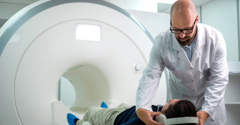
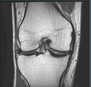
MR arthrogram uses an MRI scan after injecting the contrast material, called Gadolinium, into the joint. This contrast material also outlines the structures within the joint, same as with the other forms of arthrogram, allowing the radiologist on the cross-sectional MRI images. Unlike other imaging techniques that use X-Rays, an MRI does not expose patients to the potentially harmful effects of radiation.
An Arthrogram generally does not require any special preparation. Wear loose, comfortable clothing with easy access to the joint being examined. If it is being followed by an MRI, you should prepare the same way you would for that exam by dressing in clothes without any metal snaps or zippers.
An arthrogram is a safe procedure. As with any procedure, there are potential risks:
– An allergic reaction to the contrast dye is rare as it is injected into the joint. Please notify your physician and our technologist if you have any history of allergic reactions to Iodine.
– X-Ray radiation is part of the exam. As such, please notify our technologist if you may be pregnant to reduce potential radiation exposure.
– An arthrogram is not recommended for people with active arthritis or joint infections.

