
Many people experience pain in the lumbar spine or lower back. Often, this pain goes away on its own, but when it doesn’t, it can seriously impact your ability to go about your daily activities.
Back pain is one of the most common sources of pain that can impact our daily activities and overall mood. If you have been living with lower back pain, your doctor may order a lumbar spine MRI to help pinpoint the cause of your pain. Learn more about how to talk to your doctor about asking for an MRI to understand the cause of your back pain.
An MRI of the lumbar spine can help sort out the cause of the pain you have been experiencing, to help diagnose the source of your pain, and to help find a treatment to ease your symptoms.
In this guide, we’ll look at what a lumbar spine MRI can show, the common conditions it can help diagnose, as well as nerve conditions, and the more serious conditions a lumbar spine MRI can reveal. Let’s find out more.
What does an MRI for lower back pain show?
Lower spine pain can stem from many different issues, ranging from muscle soreness to problems with the vertebrae and the disks that separate them. An MRI scan provides detailed images of the lower back, including bones, joints, nerves, and soft tissues. Common causes include:
- Sprains and strains
- Disk problems, such as a herniated disk or degenerative disk disease
- Sciatica (nerve pain)
- Osteoarthritis
- Compression fracture of the vertebra
- Spinal stenosis
In most cases, lower back pain is not caused by a serious problem. However, any of the muscles, organs and tissues in the lumbar area can be the source of pain, which is why an accurate diagnosis is important.
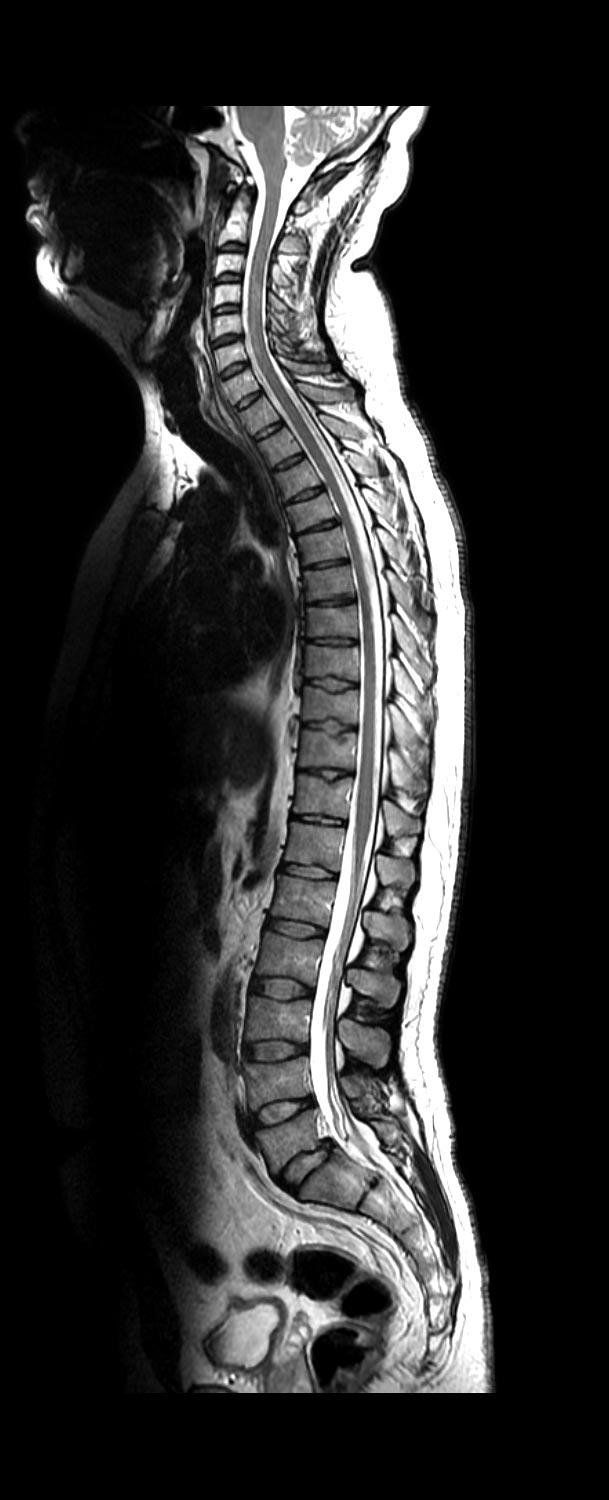
MRI imaging can be a helpful tool, as it gives a detailed view of your spinal anatomy. A lumbar spine MRI shows:
- The alignment of your spine
- Details about the intervertebral disks, such as whether they appear normal, herniated, dehydrated or worn down
- The size of your spinal canal and whether the spinal cord is being compressed
- Whether any nerves are compressed or inflamed
- Injuries to bones, ligaments and the spinal cord
- Any birth defects on or near the spine that could cause pain
- Tumors in the spinal cord, vertebrae, nerves or surrounding tissues
- Signs of infection
These images can help your healthcare provider understand more about the source of your back pain, whether it may be from an injury, structural issue, or other concern.
Your doctor will use the results of an MRI along with findings from a physical exam and your symptoms to arrive at a diagnosis and recommended treatment plan.
Touchstone Medical Imaging offers high-quality MRI imaging at up to 60% less than the cost of hospital imaging. After getting a doctor’s order, make an appointment at one of our many convenient centers.
How does an MRI scan create detailed images of your spine?
An MRI scan uses a strong magnetic field and radio waves to create images of your spine. The radio waves react to the magnetic field and create signals that are sent to a computer that reads the signals and turns them into images of the spine and surrounding tissues.
What can an MRI show about the bones, joints, and soft tissues of my back?
An MRI scan can reveal many things about the bones, joints, and soft tissues of your back, including fractures of the spine, spinal misalignment, and infections or growths that might be present. It can also show inflammation or arthritis, and soft tissue damage such as muscle strains, nerve compression, and bulging discs.
What changes related to normal aging or wear and tear can be seen on MRI results?
Normal changes that can accompany aging can be seen on MRI results. These include disc degeneration or thinning, bone spurs, arthritis, and changes in spinal stability and alignment. Sometimes these changes can be the source of lower back pain, but sometimes they are also present without significant symptoms.
What to expect when getting a lumbar MRI
An MRI is a noninvasive scan that uses magnets and radiofrequency waves to create detailed images of internal structures of the body. A low-risk procedure, MRI does not expose you to any radiation.
An outpatient MRI appointment need not take much time out of your day. A technologist will help you lie down on an exam table that slides into the MRI machine. Usually, the entire process takes about 30 to 60 minutes. After the exam, a board-certified radiologist will interpret your results, providing information to help your doctor create a treatment plan.
A convenient way to get answers to back pain
Life is busy, and it can be tempting to ignore lower back pain and simply hope it goes away. But it’s important to determine what’s causing your lumbar spine pain and ensure it’s not a sign of a more serious condition. Touchstone Medical Imaging makes getting an MRI fast and easy, with multiple centers and convenient hours, including evenings and weekends.
Touchstone Medical Imaging’s subspecialized, board-certified radiologists provide doctors with precise results they can trust. If your doctor has ordered an MRI to help diagnose back pain, make an appointment at a Touchstone Medical Imaging Center near you.

Common spine conditions an MRI can detect
An MRI scan will show healthy tissues as well as tissues that are damaged or inflamed, revealing your soft tissues and bones, which can help narrow down the sources of your back pain.
What signs of disc problems, like bulging or herniation, can an MRI show?
An MRI will show whether the discs in your lower spine are bulging, which means the inner part of the disc is wider than normal, and bulging into the spinal canal.
It can also show if a disc has been herniated, which means the inner part of the disc has ruptured or pushed through the outer layer. Both bulging discs and herniated discs can press on nerves and cause pain. An MRI will show the location and extent of disc problems in the lumbar spine.
What indications of arthritis or degenerative disc disease are visible on a lumbar spine MRI?
Signs of degenerative disc disease that are visible on a lumbar spine MRI include loss of disc hydration, which results in flattening of the discs, visible tears in the outer layer of the discs, and narrowing of the spinal canal.
Arthritis may appear as inflammation of the discs or bone spurs (small bony growths) that appear along the edges of joints. Narrowing of the spinal canal can press on nearby nerves as a result of arthritis and disc degeneration.
How does a lumbar spine MRI detect the inflammation and swelling of soft tissues?
A lumbar spine MRI will show differences between healthy soft tissues and inflamed tissues surrounding the spine. Swollen or inflamed tissue retains fluid, and this change in tissue structure appears brighter on the MRI. Inflammation of nerves will also appear brighter on the MRI.
Irritated nerves may also appear thicker on the scan. Inflamed and swollen tissues appear brighter on the MRI because the scan measures changes in water retention and changes in tissue structure. These can include changes in tissues that result from muscle strains and ligament tears.
What a lumbar spine MRI can reveal about your nerves
One reason the lower back can be a source of pain for many people is because it contains nerves that can become irritated by pressure from surrounding tissue or discs in the spine. An MRI can help show where this irritation is taking place, and what the cause or causes might be.
What evidence of nerve compression, like spinal stenosis, can an MRI scan detect?
Spinal stenosis can be seen on an MRI as a narrowing of the spinal canal on the images. This narrowing can be caused by bulging discs or arthritis, bone spurs or thickened ligaments. This reduced space makes it difficult for nerves and the spinal column to avoid being compressed, which can result in pain, weakness, or numbness.
How can an MRI show the cause of nerve irritation or inflammation in my back?
An MRI shows changes in soft tissues, and it can show if nerve irritation is caused by herniated discs or other structural issues that are pressing on the nerves. If nerves are inflamed or damaged they will appear brighter on the scan.
The presence of infection, which can also irritate nerves, will also be seen on the scan. An MRI reveals both structural changes in the spine and tissue changes surrounding the spine, both of which can cause nerves to be irritated.
What does an MRI show about conditions like sciatica or pinched nerves?
Structural changes such as herniated discs and narrowing of the spinal canal can contribute to conditions like sciatica or pinched nerves.
Sciatica is caused by pressure on the sciatic nerve, which can be seen on the scan. Nerves that are irritated by structural pressure can be seen on the scan as reacting to this pressure, whether from a bone spur or narrowing of the spinal canal.
Serious conditions a lumbar spine MRI can find
Although lower back pain is not an unusual concern, it can really affect your quality of life, and sometimes it can indicate a serious condition that needs attention. This might include unusual growths such as tumors or cysts, and also infections that might be present. A lumbar spine MRI can also reveal degenerative conditions of the spine, which can be age-related, as well as the result of an injury or your genetics.
How does a lumbar spine MRI scan detect tumors, cysts, and abnormal growths?
The magnetic field and radio wave imaging of a lumbar spine MRI will detect tumors, cysts, and other abnormal growths. These conditions will appear on the scan as different from healthy tissue, and different from one another based on tissue density and internal structure. A cyst will be filled with fluid, while a tumor is solid in nature.
How can an MRI scan show the signs of infection or inflammation?
An MRI scan reacts to infection or inflammation by showing areas of swelling or fluid buildup. When tissues are inflamed or infection is present, they hold onto water, which is seen on the scan as areas of increased signal intensity. Even subtle changes in infected or inflamed tissue can be detected by the radio wave signals that encounter these conditions.
What are the degenerative conditions a lumbar spine MRI can help to diagnose?
Degenerative conditions of the lumbar spine result from wear and tear of the spine, also called spondylosis. Conditions that can be seen on a lumbar spine MRI include herniated or bulging discs, arthritis, thinning of discs, narrowing of the spinal canal, thickening of ligaments, and bone spurs. These conditions are made clear by the images found in a lumbar spine MRI.
What to expect when getting a lumbar MRI
An MRI is a noninvasive scan that uses magnets and radiofrequency waves to create detailed images of internal structures of the body. A low-risk procedure, MRI does not expose you to any radiation.
An outpatient MRI appointment need not take much time out of your day. A technologist will help you lie down on an exam table that slides into the MRI machine. Usually, the entire process takes about 30 to 60 minutes. After the exam, a board-certified radiologist will interpret your results, providing information to help your doctor create a treatment plan.
A convenient way to get answers to back pain
Life is busy, and it can be tempting to ignore lower back pain and simply hope it goes away. But it’s important to determine what’s causing your lumbar spine pain and ensure it’s not a sign of a more serious condition. Touchstone Medical Imaging makes getting an MRI fast and easy, with multiple centers and convenient hours, including evenings and weekends.
If your doctor has ordered an MRI to help diagnose back pain, make an appointment at a Touchstone Medical Imaging Center near you.
Frequently Asked Questions for a Lumbar Spine MRI
A MRI provides detailed images of the bones, joints, discs, and soft tissues in the lower back, helping to diagnose several spine conditions.
MRI uses strong magnetic fields and radio waves to create high-resolution images of the structures in your lower back.
It can reveal fractures, joint inflammation, disc abnormalities, ligament damage, and other structural issues affecting the lower back.
Yes, it can show degenerative changes like disc wear, arthritis, and spinal narrowing, all of which can occur naturally over time.
It can detect bulging, herniated, or degenerated discs that may contribute to pain and nerve compression.
It identifies narrowed spinal canals, pinched nerves, and other abnormalities that cause pain, numbness, or weakness.
Yes, it can identify abnormal growths, swelling, or signs of infection that may require further medical evaluation.
It can confirm conditions like degenerative disc disease, osteoarthritis, and spondylosis, which affect spinal stability and function.