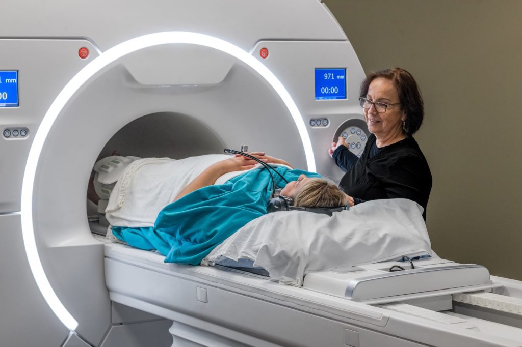
If you’re experiencing back pain or leg pain, or numbness in your back and legs, your healthcare provider may have suggested a lumbar spine MRI to get a closer look at your spine.
A lumbar spine MRI is one of the most effective tools for diagnosing herniated discs because it provides highly detailed images of the structures in your lower back, like your spine’s discs, vertebrae, and nerves.
We’ll show you why a lumbar spine MRI is the preferred scan for diagnosing herniated discs, how an MRI shows herniated discs, and how an MRI can help diagnose sciatica and spinal stenosis.
How does an MRI show detailed images of my back’s discs, vertebrae, and nerves?
An MRI scan uses strong magnets and radio waves to create detailed, cross-sectional images of your spine. When it comes to diagnosing herniated discs, an MRI is incredibly useful because it can show the condition of the discs between your vertebrae, along with the surrounding nerves.
MRI scans can clearly differentiate between bone and soft tissue, which means it can reveal if a disc has slipped, or if a disc is pressing on a nerve. This level of detail can help your healthcare provider to pinpoint exactly where the problem is, and how severe it might be.
What makes MRI the best option for imaging soft tissue issues like herniated discs?
One reason an MRI is considered the gold standard for diagnosing herniated discs is its ability to produce high-resolution images of soft tissue. Your spine’s discs, which are made of soft, gel-like material, are difficult to see clearly using other types of scans.
An MRI provides clear pictures of the spinal discs, making it easier for your healthcare provider to see if any disc material is bulging, or has broken through the outer layer. It also shows if the discs are impacting nearby nerves and spinal structures, which is very important for diagnosing a condition like herniated discs.
How can an MRI help detect subtle problems that may not show on other scans?
In many cases, herniated discs can cause subtle changes in your spine that are hard to detect without a thorough scan. These might include slight disc bulges, small tears in the disc, or mild nerve compression. An MRI can pick up on these subtle issues that might otherwise go unnoticed.
This level of detail is especially important if you’ve been dealing with symptoms for a while but haven’t yet received a clear diagnosis. An MRI can help identify the root cause of your discomfort, allowing you and your healthcare provider to develop a targeted treatment plan, so you can get some relief from back pain.
Understanding herniated discs
Herniated discs can cause a variety of symptoms, not only back pain and discomfort, but also pain that radiates down your legs. A lumbar spine MRI is often recommended to get a clearer picture of whether you have a herniated disc in your back.
Why are spinal discs important? What do they do?
Spinal discs act like cushions between the bones of your spine, or vertebrae. Each disc has a soft, gel-like center and a tougher outer layer. They help absorb the shock and pressure your spine experiences when you move, lift, or twist.
Together, they keep your spine flexible and allow for smooth movement, while also protecting the bones in your spine from grinding against each other. Without healthy discs, your spine would lose much of its ability to move fluidly, which can lead to back pain and stiffness.
How can a herniated disc impact the surrounding vertebrae and nerves?
When a disc becomes herniated, it means that part of the soft inner material has pushed through a tear or weakened spot in the outer layer. This bulging material can press on nearby nerves, or even the spinal cord itself, leading to a range of symptoms. You might feel sharp or shooting pain, tingling, numbness, or muscle weakness.
If the herniation is large enough, it can also affect the vertebrae, causing inflammation and irritation around the joints in your spine. In some cases, a herniated disc can even cause the vertebrae to shift slightly, which can further compress nerves and make the pain worse.
How does an MRI show if a herniated disc is impacting my vertebrae and nerves?
An MRI is an excellent tool for detecting herniated discs because it provides a clear image of both the discs and the surrounding vertebrae and nerves. The scan can reveal exactly where the disc material is pressing on a nerve or how it’s affecting the vertebrae. This level of detail allows your healthcare provider to see how severe the herniation is and whether any nerve roots are being compressed.

Diagnosing herniated discs with MRI scans
A lumbar spine MRI scan is valuable because it can reveal the exact location, size, and severity of the herniated disc, along with any nerve involvement that could be causing your symptoms. Let’s see how your provider uses an MRI scan to diagnose a herniated disc.
How does an MRI help pinpoint the exact location of a herniated disc?
One of the biggest advantages of an MRI scan is its ability to show a clear, precise image of the entire lumbar spine, including all the discs and surrounding structures. This makes it easier for your healthcare provider to identify the specific disc that is herniated. An MRI produces detailed images from different angles, allowing your provider to see exactly where the disc has slipped or bulged.
How can an MRI scan reveal the size and severity of the herniation?
In addition to pinpointing the location, an MRI can also show how large the herniation is, and how far the disc material has pushed out from its normal position. This is important because the size of the herniation often affects the severity of your symptoms.
A small bulge might cause mild discomfort, while a more significant herniation can put more pressure on the surrounding nerves, leading to more intense pain or numbness. With an MRI, your healthcare provider can assess the extent of the damage, and determine how much of the disc has herniated.
How does an MRI identify the nerve compression or irritation from a herniated disc?
Herniated discs often press against nearby nerves, leading to symptoms like pain, tingling, or weakness in the legs. An MRI is particularly useful for detecting this nerve compression or irritation because it provides a clear view of the soft tissues, including the nerves themselves.
MRIs can show whether a herniated disc is pressing directly on a nerve root, or causing inflammation around the nerves, which is important for understanding why you’re experiencing certain symptoms.
Evaluating conditions that herniated discs can cause: sciatica and spinal stenosis
A lumbar spine MRI is not only useful for diagnosing herniated discs, but also for identifying conditions caused by herniation, like sciatica and spinal stenosis. These conditions can lead to significant pain and discomfort, and by providing detailed images of your spine, an MRI can help your provider determine whether a herniated disc is the source of your pain.
How can a lumbar spine MRI show if a herniated disc is causing sciatica or other nerve pain?
Sciatica happens when a herniated disc compresses the sciatic nerve, which runs from your lower back down through your legs. It can cause sharp pain, tingling, or numbness that radiates along the path of the nerve. A lumbar spine MRI is excellent for revealing whether a disc is pressing on the sciatic nerve or other nearby nerves.
That’s because an MRI scan gives your provider a close look at the herniated disc, and at whether it’s near any nerves, helping to pinpoint the exact cause of your nerve pain. With this information, your healthcare provider can confirm whether your symptoms are due to sciatica, or to another form of nerve irritation.
How does an MRI scan show spinal stenosis and inflammation around the disc?
Spinal stenosis is when the space around your spinal cord and nerves becomes narrowed, often because of inflammation, or a bulging disc pressing into the spinal canal. This narrowing can cause pain, numbness, or weakness, especially when you’re standing or walking.
A lumbar spine MRI can clearly show whether a herniated disc is causing this narrowing, as well as any inflammation that may be contributing to your symptoms. By highlighting these structural changes in your spine, an MRI allows your healthcare provider to understand the severity of the stenosis, and how it’s impacting your spinal cord and nerves.
How will my MRI results help my healthcare provider to create a treatment plan?
Once your MRI results are available, your healthcare provider will have a comprehensive view of your spine and a radiology report analyzing the images from a subspecialized radiologist, including any herniated discs, and related conditions like sciatica or spinal stenosis.
With this information, they can develop a treatment plan tailored specifically to your condition, whether your treatment involves physical therapy, medication, or surgery. Your MRI results will provide the clarity your provider needs to make the best decision for your care.

How to schedule your MRI appointment with us
Touchstone Medical Imaging offers MRI scans in Arkansas, Colorado, Florida, Montana, Oklahoma, and Texas.
Reach out to us at Touchstone, and we’ll help you schedule an CT appointment at an imaging center near you, today.
We’re here to help you get the answers you need.
Frequently Asked Questions (FAQ)
An MRI produces high-quality images that give a clear picture of your discs, vertebrae, and nerves, helping your health provider to spot any irregularities.
MRI scans are excellent for showing soft tissue structures like discs and nerves, giving more detailed information than other types of imaging.
Yes, an MRI can uncover subtle issues in the spine, like minor herniations or nerve problems that might not appear on other scans.
Spinal discs serve as cushions between the vertebrae, helping you to move around, and absorbing any impacts to the spine.
A herniated disc can press on nearby nerves or the spinal cord, which may lead to symptoms like pain, numbness, or weakness.
MRI scans can clearly display the interactions between a herniated disc and the surrounding nerves or vertebrae, showing any compression or irritation.
Yes, an MRI can reveal whether a herniated disc is putting pressure on nerves like the sciatic nerve, which can cause back pain.
The information from an MRI allows your doctor to better understand the location and severity of the herniation, which helps guide treatment options.

