
When people find out they need a brain MRI, some of them start to worry. We completely understand why.
It’s always OK to be concerned about your health, but a brain MRI is nothing to worry about.
If your healthcare provider has recommended that you get a brain MRI, it just means they want to get a closer look at your brain, and they think an MRI would be the best scan for you. That’s it.
Let’s learn more about what you can expect from a brain MRI, so you can see what they’re really like.
We’re going to take a close look at why your provider recommended a brain MRI, at what brain MRIs show, and at what you can expect from your results.
What is an MRI and how does it work?
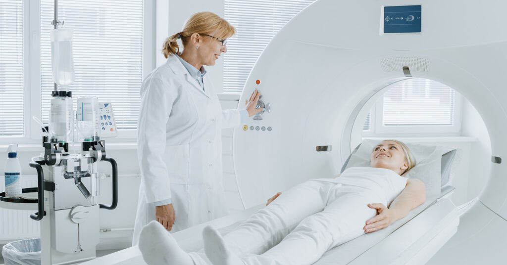
MRI stands for Magnetic Resonance Imaging. It’s a non-invasive diagnostic tool, which means that it allows doctors to see inside your body without using any incisions.
Instead of using radiation, MRIs harness the power of magnetic fields and radio waves to create detailed images of the soft tissues within your body.
The MRI machine captures signals from your body, and with the help of a computer, converts those signals into detailed images for the radiologist to analyze.
The result is clear, intricate pictures of your brain’s structure and tissues.
How is a brain MRI different from other types of MRI scans?
A brain MRI focuses primarily on the brain and head and, sometimes, on the surrounding structures like the spinal cord.
What makes the brain MRI special is the level of detail it provides about the brain’s soft tissues, such as gray matter, white matter, blood vessels, and fluid compartments.
This detail enables healthcare providers to diagnose and monitor a range of neurological conditions, from tumors and inflammations to developmental anomalies and degenerative disorders.
How safe is a brain MRI?
A brain MRI is considered one of the safest imaging techniques available. Since MRIs don’t use radiation, you won’t have to worry about radiation exposure.
However, because of the strong magnets used in the procedure, it’s important to let your healthcare provider know if you have any metal implants or devices inside your body.
These can interact with the magnetic field and might pose safety risks or distort the images.
As a rule of thumb, if you’re unsure about anything before the procedure, always provide please ask questions. We’re happy to help.
How long does a typical brain MRI procedure take?
The duration of a brain MRI can vary based on what exactly your healthcare provider is looking for, so be sure to plan accordingly.
On average, a typical brain MRI lasts between 30 to 60 minutes. Sometimes, contrast agents might be used to enhance certain images, which can add a bit of time.

Why your healthcare provider recommended a brain MRI
Now that we understand what a brain MRI is, let’s see why your healthcare provider thinks a brain MRI is the right choice for your healthcare.
MRI is a unique kind of imaging technique that can see and do things that other imaging techniques, like CT scans, cannot see or do.
How does an MRI differ from other imaging techniques?
Unlike some other imaging methods, MRIs don’t rely on radiation. This makes them particularly useful for capturing in-depth images of soft tissues, like those found in the brain.
The pictures produced by an MRI offer a detailed and clear view of the brain’s anatomy.
These images can showcase subtle differences in tissue health and structure, making it easier for healthcare providers to spot abnormalities or potential issues that other imaging methods might miss.
What symptoms or conditions might prompt a doctor to recommend a brain MRI?
There’s a range of symptoms and conditions that could lead your healthcare provider to suggest a brain MRI.
Some of these include:
- Persistent headaches or migraines that don’t have an apparent cause
- Unexplained dizziness or episodes of vertigo
- Seizure activity or sudden changes in behavior
- Memory problems or cognitive changes that can’t be explained by other factors
- Visual disturbances or loss of vision
- Weakness or numbness in certain parts of the body
- Suspicion of certain conditions like multiple sclerosis, tumors, inflammations, or aneurysms
Can an MRI detect changes in the brain before symptoms appear?
Yes, one of the most significant advantages of a brain MRI is its ability to detect changes in the brain even before symptoms manifest.
This is particularly crucial for conditions where early detection can make a substantial difference in treatment outcomes.
For example, certain types of tumors or growths might not present any symptoms until they’ve reached a particular size. With an MRI, these can be identified at an earlier stage, allowing for more timely interventions.
Similarly, degenerative diseases or conditions that affect blood flow might show early changes on an MRI before any clinical signs become apparent.
Is a brain MRI used for both diagnostic and follow-up purposes?
Yes, absolutely: a brain MRI is a versatile tool in the medical world.
Initially, it can be used for diagnostic purposes, helping healthcare providers pinpoint the cause of your symptoms or validate suspicions of certain conditions.
Once a diagnosis is made, a brain MRI can be used for follow-up purposes to:
- Monitor the progression of a condition
- Assess the effectiveness of treatments or interventions
- Check for any potential complications or new developments
If you’ve had a brain MRI before and are recommended to have another one, it could be part of a routine follow-up, or to gain deeper insights into your health status.
What a brain MRI can show
Healthcare providers choose brain MRIs because the images are so clear, detailed, and informative.
Your healthcare team will study these images to make sure your brain looks healthy.
When your provider looks at your MRI results, they’re looking at the intricate anatomy of your brain, in great detail.
What can an MRI show my doctor?
An MRI provides a close-up view of the brain’s intricate structures.
Here are some of the main parts of the brain it can highlight:
- Cerebral hemispheres: These are the large, outermost parts of the brain responsible for thinking, decision-making, and sensory processing
- Cerebellum: Situated at the back of the brain, the cerebellum plays a key role in coordination and balance
- Brainstem: Acting as a bridge between the spinal cord and the brain, the brainstem controls many vital functions, like breathing and heart rate
- Lobes of the brain: Including the frontal, parietal, temporal, and occipital lobes, each responsible for different functions ranging from motor skills and speech to vision and hearing
- Ventricles: These are fluid-filled cavities in the brain. They play an essential role in cushioning the brain and removing waste
How detailed are the images captured by a brain MRI?
Using the combined power of magnetic fields and radio waves, MRIs can produce high-resolution pictures of the brain’s soft tissues.
This allows healthcare providers to differentiate between different types of tissues, such as gray matter, white matter, and even various types of fluid in the brain.
Whether it’s a tiny blood vessel, a minute growth, or subtle changes in the brain tissue, a brain MRI is adept at highlighting these intricate details.
What do the different shades and patterns mean on a brain MRI?
On MRI images, various shades of gray represent different tissues and structures.
On your MRI results, darker shades can represent fluid-filled areas (like cerebrospinal fluid), medium grays often depict softer tissues (like gray matter), and lighter shades depict denser tissues (like white matter or large blood vessels).
Occasionally, if your MRI requires a contrast agent, areas of concern or specific structures might appear brighter to make them stand out more.
Different patterns can indicate various conditions or anomalies. For example, a cluster or spot might suggest a growth or tumor, while a pattern resembling a lesion or scarring might be indicative of conditions like multiple sclerosis.
Your radiologist is trained to interpret the patterns, shades, and nuances in order to provide insights into the health and functioning of your brain.
How a brain MRI helps your healthcare provider make a diagnosis
The images from a brain MRI are detailed enough to show when the tissues of the brain don’t look as expected.
An MRI of the brain can find abnormal growths, or an infection, and inflamed tissue. It can also diagnose brain injuries, and degenerative diseases, like Alzheimer’s or Parkinson’s.
One of the reasons why providers choose MRI scans is because they can help diagnose lots of different health conditions.
How does an MRI help in detecting tumors or growths in the brain?
MRI scans are particularly effective in pinpointing tumors or growths within the brain.
The high-resolution images they produce are capable of differentiating between the brain’s soft tissues, enabling them to identify abnormal growths even if they’re surrounded by healthy tissue.
To spot certain types of growths, a contrast agent can accentuate specific areas of the brain, especially those with increased blood flow often found in many tumors.
Importantly, tumors can have varying appearances; some might appear solid, while others are filled with fluid. The MRI provides clarity on these differences, aiding in both diagnosis and understanding the tumor’s nature.
How effective is an MRI in spotting infections or inflammations in the brain?
MRI scans play a pivotal role when identifying infections or inflammations in the brain, like meningitis or encephalitis.
If a contrast agent is used during the MRI, then the affected areas might appear more prominently, signaling a possible concern.
Moreover, any infection or inflammation can induce swelling or shifts in tissue characteristics. MRIs, with their detailed imaging, can discern these changes, setting them apart from normal brain tissue.
Can an MRI diagnose brain injuries?
MRIs are vital instruments in diagnosing brain injuries.
Although some injuries, such as concussions, might not show obvious structural changes, an MRI can detect minor abnormalities that could be overlooked by other methods.
MRIs can also effectively capture contusions (or bruises of the brain), providing insights into where the injury is, its size, and the degree of its severity.
How does MRI help detect degenerative conditions like Alzheimer’s or Parkinson’s?
While diseases like Alzheimer’s or Parkinson’s typically require clinical evaluations for primary diagnosis, MRIs are invaluable companions in the diagnostic journey.
Degenerative conditions can cause specific regions of the brain to shrink, a phenomenon known as brain atrophy. An MRI can detect this, especially during the early stages when changes might be barely noticeable.
Different diseases impact various parts of the brain: Parkinson’s, for instance, might affect areas tied to movement, while Alzheimer’s could influence regions linked to memory.
The symptoms of these degenerative diseases can mimic other conditions. In that case, an MRI helps to exclude other potential causes like tumors or strokes, and ensures the most accurate diagnosis possible.
Your brain MRI results
Your provider ordered a brain MRI because of the results you’ll get, afterwards.
No matter what they show, your imaging results will show your provider what to do next, if any further care is necessary.
Let’s see what you can expect from your brain MRI results, including what you can expect if anything looks abnormal.
How do radiologists interpret the findings from a brain MRI?
When radiologists look at your brain MRI, they’re searching for contrasts between different tissues, any abnormal patterns, or structures that don’t seem typical.
Using their extensive knowledge of the brain’s anatomy and various medical conditions, they piece together the story these images are telling.
By comparing the MRI images to what’s considered typical, and noting any differences, they can identify any concerns or give an all-clear signal.
How soon can patients expect their results after the MRI?
Generally, after the MRI is completed, the images are sent to a radiologist for review.
Depending on the complexity of the images and the clinic’s workflow, you can typically expect your results within a few days to a week.
However, if your healthcare provider is particularly concerned about a specific issue, they might expedite the review process, and you could get your results sooner.
What happens if my brain MRI results show an abnormality?
It’s essential to remember that abnormality doesn’t automatically mean something severe. The brain is complex, and there are numerous reasons an MRI might show something out of the ordinary.
If an issue is detected, your healthcare provider will discuss the findings with you in detail.
They’ll explain the nature of the abnormality, whether it’s a structural change, a growth, or another type of issue.
Your provider will answer any questions you have, ensuring you fully understand the findings and what they might mean for your health.
What are the potential follow-up steps or additional tests after a brain MRI?
If your MRI results necessitate further investigation, there are several paths your healthcare journey might take:
- Follow-up MRI: Sometimes, for clarity or additional information, another MRI might be scheduled, possibly with a different kind of contrast agent to highlight certain areas
- Other imaging studies: Depending on the findings, other imaging modalities like a CT scan or a PET scan might be suggested to provide different types of visual information
- Biopsy: If a growth is detected, a biopsy might be recommended to determine its nature—whether it’s benign (non-cancerous) or malignant (cancerous)
- Referral to a specialist: You might be referred to a neurologist or another specialist for a more in-depth assessment based on your MRI results
- Treatment plan: If a specific condition is diagnosed, your healthcare provider will discuss treatment options, whether it’s medication, therapy, or other interventions
Remember that you’re not alone in this. Your healthcare team is there to guide you, answer questions, and ensure you have a clear understanding of the road ahead.
How to schedule a brain MRI appointment with us
Touchstone Medical Imaging offers MRI imaging in Arkansas, Colorado, Florida, Montana, Oklahoma, and Texas.
Reach out to us at Touchstone, and we’ll help you schedule an appointment at an imaging center near you, today.
Did you know our MRIs cost up to 60% less than what you pay at a hospital?
We’re here to help you get the answers you need.

Frequently Asked Questions About a Brain MRI
An MRI, or Magnetic Resonance Imaging, is a medical imaging technique that uses magnetic fields and radio waves to capture detailed images of the body’s internal structures.
While X-rays and CT scans use ionizing radiation, an MRI uses magnetic fields and radio waves, offering detailed images without radiation exposure.
Providers might recommend a brain MRI for symptoms like persistent headaches, seizures, or unexplained neurological symptoms, and to investigate conditions like tumors or multiple sclerosis.
An MRI can highlight various parts of the brain, including the cerebral cortex, cerebellum, brainstem, and the deep brain structures like the basal ganglia and thalamus.
An MRI provides high-resolution images that can reveal abnormal growths, tumors, or cysts in the brain by highlighting differences in tissue characteristics.
Radiologists analyze the MRI images, looking for abnormalities in brain structures and tissue characteristics to make a diagnosis.
If an abnormality is found, doctors may recommend further tests, consultations with specialists, or a treatment plan based on the MRI findings.