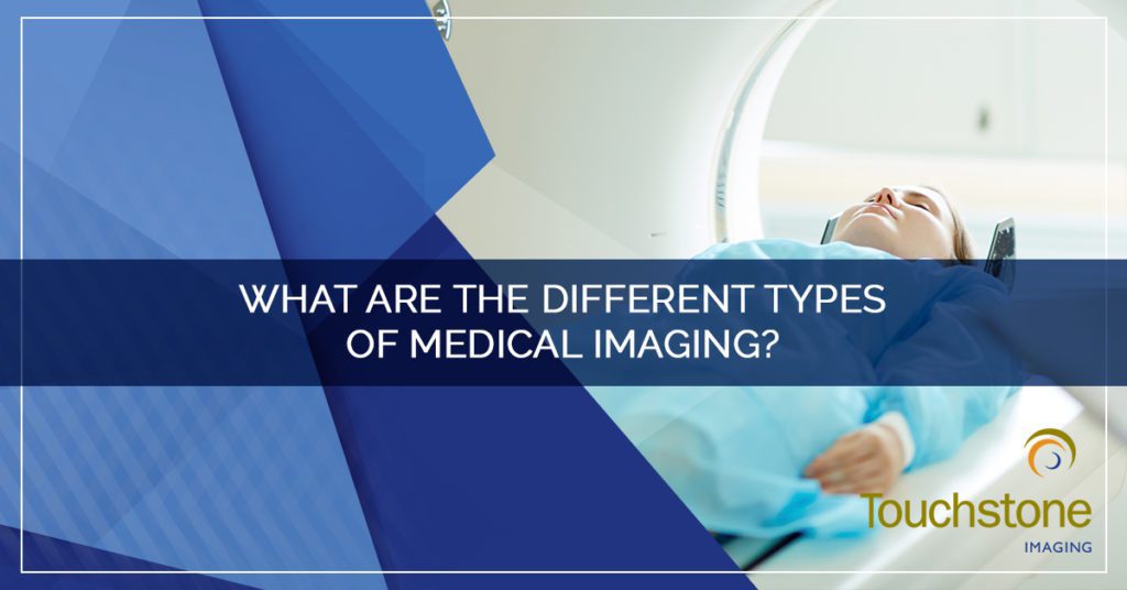
Welcome to Touchstone Imaging! We are the nation’s leading provider of diagnostic imaging services and operate medical imaging facilities all over the country. We offer a wide range of diagnostic imaging services to our patients. In this article, we’ll outline the various types of medical imaging services that we offer and the differences between them. We hope this helps you know more about your upcoming exam or procedure, or simply stay educated regarding a potential procedure.
MRI
An MRI, or magnetic resonance imaging, is a painless way that medical professionals can look inside the body to see your organs and other body tissues. It’s designed to detect injured tissue or potentially harmful changes in your body. The MRI uses a large magnet, through radio waves, and a computer to generate images of your organs and tissues. There is no radiation involved with an MRI, and there are no known side effects.
During your MRI, you will lay down on a padded table which slides into the MRI machine. The procedure will last approximately 30 minutes to an hour, depending on what body area is being scanned, and it’s important to lie as still as you can. If you experience any discomfort, it’s important to tell the technician performing your scan.
CT Scan
A CT scan is also known as a CAT scan, or computed tomography scan. It is a special kind of X-ray that takes a picture of a cross-section of a specific part of your body. The purpose of a CT scan is to find specific changes inside your body that a normal X-ray machine may not be able to find. A CT scanner is a large X-ray tube that moves around your body and sends signals to a computer that then creates an image. The average CT scan takes approximately five to 10 minutes.
PET/CT
A PET/CT scan, or positron emission tomography, is an exam that uses a small amount of radioactive material to reveal the function of an organ or tissue. It is used to detect disease that begins with functional changes. When you arrive for your PET/CT scan, a technologist will then insert a catheter into a vein in your arm and inject a radiotracer. The entire procedure takes approximately an hour plus an additional 20 to 30 minutes for the images to be completed.
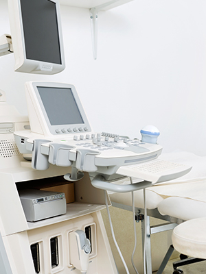 Ultrasound
An ultrasound uses sound waves to produce images of the inside of the body. It is safe and painless, utilizing a small probe and gel placed directly on the skin. High-frequency sound waves are transmitted from the probe, through the gel, and into the body. A computer is connected to the probe and uses the sound waves to create an image. Ultrasounds do not use radiation, so there is no risk of exposure.
X-Ray
An X-ray is the oldest form of medical imaging. It is a painless, non-invasive procedure that helps medical professionals diagnose and treat medical conditions. Radiography involves exposing a part of the body to a small amount of radiation to produce a picture of that body part below the skin. It is often used to diagnose broken bones, infections, injury, or even locating foreign objects within soft tissue.
Arthrogram
An arthrogram is a diagnostic imaging procedure that uses X-rays to guide and evaluate the injection of contrast directly into a joint. The purpose of an arthrogram is to get more information about joints than a more traditional medical imaging procedure, like an MRI or CT scan, can provide alone. Typically, after an arthrogram, it is required that you have an MRI or CT scan to obtain more information.
Myelogram
The purpose of a myelogram is to get more information about your joints than what a CT scan or MRI can provide. Myelograms typically find tumors, infections, problems with the spine, or problems caused by arthritis. A myelogram uses X-rays and a special dye to make pictures of the bones and fluid-filled space between bones. To get more information, a CT scan is often performed after the X-rays while the dye is still in the body.
Women’s Imaging
Women’s imaging is a broad term referring to a variety of diagnostic imaging services directly related to the needs of women. These include:
- Mammography – A mammography uses low-energy X-rays to examine the breast tissue. It is used for early detection of breast cancer.
- Screening Mammography – A screening mammogram is an X-ray of the breast tissue that is used for women who have no symptoms or signs of breast cancer. It is used to detect breast cancer when it is too small to be felt.
- Diagnostic Mammography – A diagnostic mammography is for a woman with symptoms in the breast tissue, such as a lump.
- 3D Mammography – 3D mammography is used for early detection of breast cancer. It uses new imaging technology to detect tumors sooner.
- Breast Ultrasound – A breast ultrasound is used for taking a closer look at areas of breast tissue following a mammogram.
- DEXA Bone Density – A DEXA bone density scan is an enhanced form of X-ray technology that is used to measure bone density loss.
- Breast MRI – A breast MRI uses a computer, magnet, and radio waves to produce images of breast tissue to evaluate for potential risks.
At Touchstone Imaging, we are committed to quality, compassionate care. We offer patients high-quality outpatient imaging services and support them with honesty about their procedure. Find a Touchstone Imaging location near you.