When most people think of medical imaging, they think of the diagnostic purposes it serves — identifying a tumor, visualizing a broken bone, or searching for a strain or sprain. While diagnostic imaging is a very important factor in diagnosing and treating ailments, medical imaging is also useful in other medical applications. In today’s post, we are going to explore some of the ways medical imaging can be used to verify accuracy in treatments and procedures.
At Touchstone Imaging, we offer a variety of medical imaging modalities that can be used for several purposes. Whether your provider has ordered an MRI to look at a torn ligament or you need a follow-up x-ray to visualize a healing fracture, we can accommodate your needs.
Cancer Treatment
Patients with suspected cancer are likely to have several diagnostic imaging procedures performed. If there is confirmation of cancer, specialized imaging tests may be performed to establish a baseline before treatment. Once treatment is underway, patients may get periodic scans to ensure the treatment is effective. Whether the patient undergoes surgical removal of a tumor, chemotherapy, or radiation treatment, scans will be utilized to acknowledge various stages of cancer in the body. Repeat scans may be conducted to confirm remission and then periodically to assess for recurrent cancer.

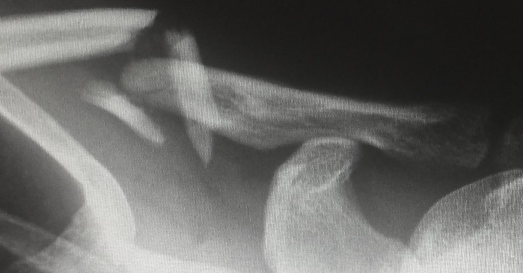
Bone Repair
In the event of a broken bone, the patient will likely have several x-rays performed throughout treatment. The initial scan will verify the broken bone and tell the orthopedist what kind of break it is, precise location, and establish an effective treatment plan. Sometimes a simple cast for a few weeks is enough, while in other cases, surgical intervention may be necessary. In either event, once treatment is complete, patients will likely undergo a final x-ray to confirm that the bone is healed before you are released to resume life as normal. This helps prevent reinjury or complications with new hardware.
Invasive Line Insertion
Central line and ports are inserted in a simple surgical procedure to avoid using distal blood vessels for long term IV use or when administering caustic medications including chemotherapy or antibiotics. Mickey buttons, ports, and gastric tubes are all externally placed access points for internal organs. Nasogastric tubes and endotracheal tubes are devices inserted through the mouth or nose to feed or ventilate. Before any of the aforementioned devices can be used for their intended purpose after insertion, placement must be verified via medical imaging. Except for an intubation tube, which will be used right away and confirmed while in use. Verifying placement with imaging scans helps to ensure that the device or tube made it to its intended destination and can be used safely.
These are all just a few examples of common reasons that medical imaging is necessary for much more than the diagnosis. Imaging technology has been used to guide treatment and verify the accuracy of interventions. For all of your medical imaging needs, find a Touchstone Imaging Center near you and schedule your appointment today.