
Historically, a diagnosis of cancer was considered a death sentence. Cancer was a word that invoked fear and panic. In fact, in 1971, the federal government declared a “War on Cancer,” which allowed for increased funding of research programs and public health programs to bring awareness to prevention and detection methods. The great news is that in the 1970s, the five-year survival rate for cancer diagnosis was less than 50 percent, while today it is nearly 70 percent. Screening and diagnostic tools have helped to increase early detection which has more favorable outcomes. Advancements and the increase in access to high-quality medical imaging have made a significant difference.
Follow along today as we review the most common and the deadliest cancers, and how medical imaging tests at Touchstone Imaging can help.
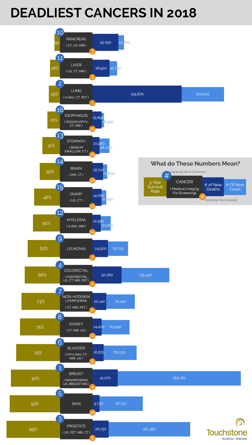
When we talk about deadliest cancers, it is important to distinguish between “deadliest” based on raw numbers of deaths the type of cancer caused and the five-year survival rate. For instance, the seven deadliest cancers based on the number of lives claimed last year are:
- Lung: 155,870
- Colorectal: 50,260
- Pancreas: 43,090
- Breast: 41,070
- Liver: 28,920
- Prostate: 26,730
- Leukemia: 24,500
However, there is a 99 percent five-year survival rate for prostate cancer and a 91 percent five-year survival rate for those diagnosed with breast cancer. Although the number of deaths are significantly higher than other cancer types, the outcomes are often much better. Based on mortality rates, the seven deadliest cancers are:
- Pancreas: 9% survival rate, 91% mortality rate
- Liver: 18% survival rate, 82% mortality rate
- Lung: 19% survival rate, 81% mortality rate
- Esophagus: 21% survival rate, 79% mortality rate
- Stomach: 31% survival rate, 69% mortality rate
- Brain: 35% survival rate, 65% mortality rate
- Ovary: 46% survival rate, 54% mortality rate
To really grasp how significant these improvements are, we can compare the numbers to 10 years ago.
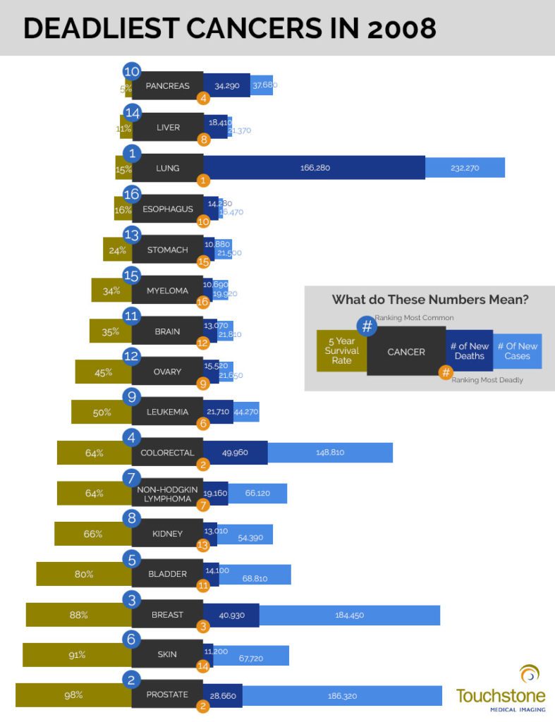
One thing you may notice in these infographics is how over the 10-year period, there was an improvement in the survival rate for every type of cancer, and two graduated off of the “deadliest cancers list,” dropping the number of cancers that have a mortality rate of more than 50 percent from nine, to seven.
The rates of breast cancer went up over the 10-year period, as did the five-year survival rate. Awareness and emphasis have been placed on early identification including self-breast exams and advanced medical imaging technology. Medical imaging has identified more cases of Stage I breast cancer that was successfully treated.
Lung cancer rates decreased while survival rates increased, indicating that anti-tobacco campaigns and reduced exposure to cigarette smoke, either by primary or secondhand methods, are working. As tobacco use rates continue to decrease, so does the incidence of lung cancer.
Increasing the number of cancer survivors with medical imaging.
The best treatment for cancer is prevention and the best outcomes derive from early identification and intervention. Medical imaging as a diagnostic tool plays an important role in the detection, diagnosis, staging, treatment, and monitoring of cancer. Let’s take a look at how each imaging modality contributes to the “War on Cancer.”
Medical imaging tests are used in many ways in cancer including:
- Screening and cancer detection
- Identification of masses
- Staging cancer progression and metastasis
- Planning radiation therapy focus
- Monitoring progression or regression during treatment
- Identifying recurrence or verify remission
A radiologist technologist will conduct the diagnostic imaging procedure, then a radiologist will interpret the results. An oncologist will use the results to diagnose and create a cancer treatment plan.
X-ray
X-rays can help detect cancer in a variety of body structures including bones, stomach, kidneys, and lungs. The benefits of x-rays are that they are less expensive, quicker, and require less preparation than other medical imaging procedures. Special x-rays include a barium enema, used to view the lower GI system, and an intravenous pyelogram (IVP), that can better visualize the urinary system.
X-ray technology can be used to detect cancer of the lung, stomach, myeloma, and bladder.
Ultrasound
Ultrasound is a useful diagnostic tool for examining soft tissues that don’t show up well on x-rays. Ultrasound can also be useful in distinguishing between fluid-filled cysts and solid tumors. Ultrasounds can also be used to see blood flow in real time, and can be used to guide a biopsy.
Ultrasounds can be used in the detection of cancers of the pancreas, liver, ovary, colorectal, kidney, bladder, breast, and prostate.
CT Scan
Computed tomography (CT) scans can be used to show a cross-section of body parts including bones, organs, and soft tissues more clearly than a standard x-ray. This can help identify masses or tumors and can show what blood vessels feed the tumor and what other structures it affects. If a tumor is identified, a CT can also be used to guide a biopsy or radiofrequency ablation as treatment for a tumor.
Recently, radiologists have been using CT technology to perform “virtual endoscopy,” which is a bronchoscopy or colonoscopy without actually having to put a scope into the body.
CT scans can be used to detect cancer of the pancreas, liver, lung, esophagus, stomach, brain, ovary, colorectal, non-Hodgkin lymphoma, kidney, bladder, and prostate.
MRI
Magnetic resonance imaging (MRI) is a powerful diagnostic tool for identifying and guiding cancer treatments. MRI takes multiple cross-sectional images from many angles to create a picture of the entire body or body part. MRIs offer much clearer images than other medical imaging modalities, and can even help doctors identify whether or not a mass is cancerous. MRI is also very useful in identifying whether cancer has metastasized to another part of the body.
MRI can be used to detect cancer of the prostate, pancreas, liver, esophagus, brain, myeloma, colorectal, non-Hodgkin lymphoma, kidney, bladder, and breast. An MRI with contrast is the best way to visualize the brain and spinal cord tumors.
Breast MRIs can be used to supplement mammography or ultrasound. It is often used to determine the extent of newly diagnosed breast cancer and can help identify metastasis. It can also be used to evaluate the effectiveness of chemotherapy and breast implants.
Bone Scan
Bone scans are used to look for cancers that may have spread to bones. For instance, prostate cancer often metastasizes to bones first, and a bone scan may help detect this.
PET Scan
Positron emission tomography (PET) scans use a form of radioactive sugar to identify cancer cells and help determine how quicking they are growing. PET scans can be used if your doctor thinks that cancer may have spread, but is not sure where.
PET scans can be used to detect the presence and metastasis of lung, colorectal, non-Hodgkin lymphoma, and prostate cancer.
At Touchstone Imaging, all of our imaging centers use state-of-the-art technology and the most current practices to produce high-quality diagnostic images to help improve the detection, diagnosis, treatment, and management of cancer. If you need medical imaging for cancer screening, visit us online to find a Touchstone Imaging center near you.

For more information, visit these online resources.