Your body is an amazing entity made up of all sorts of tissues and organs that allow you to function like a well-oiled machine. When something goes wrong with one of these parts it can cause excruciating pain that alerts you to the problem and reminds you not to use it while it recovers. When it comes to musculoskeletal injuries, it is not always easy to tell what the actual injury is or what tissue is affected, but that is important information to know because it will guide the treatment and healing process. The technology of medical imaging allows your medical provider to take a look inside your body to identify the exact location and cause of your pain as well as determine the extent of the damage. This helps your provider create a treatment plan that optimizes healing and offers the best chances of a full recovery.
At Touchstone Imaging Centers, our medical imaging facilities offer a wide range of diagnostic imaging services including x-ray, CT, and MRI to help you and your provider visualize your injury. High-quality diagnostic images are the key to accurate diagnosis and effective treatment. Join us in today’s post as we discuss some of the most common musculoskeletal injuries – sprains, strains, dislocations, and breaks – and how medical imaging helps reveal them.
Identifying Sprains
Ligaments are the fibrous tissue that connects joints to bones and helps provide support and stability for the joint. When the joint is rolled, twisted, or forcefully moved in a way the joint is not meant to move, it can cause tearing, stretching, or detachment of the ligament. This causes significant swelling and pain in the joint as well as limits the range of motion. You can sprain just about any joint and can sustain multiple sprains in the same joint if multiple ligaments are affected. Treatment for sprains will vary depending on the severity of the stretching or tearing of the ligament and usually involves ice, rest, and immobilization along with anti-inflammatories and therapeutic movements such as stretching. Sprains can take quite a while to heal because of the dense tissues that make up ligaments. It is important not to reinjure the ligament during the healing process. Ligament detachment will require surgical intervention.
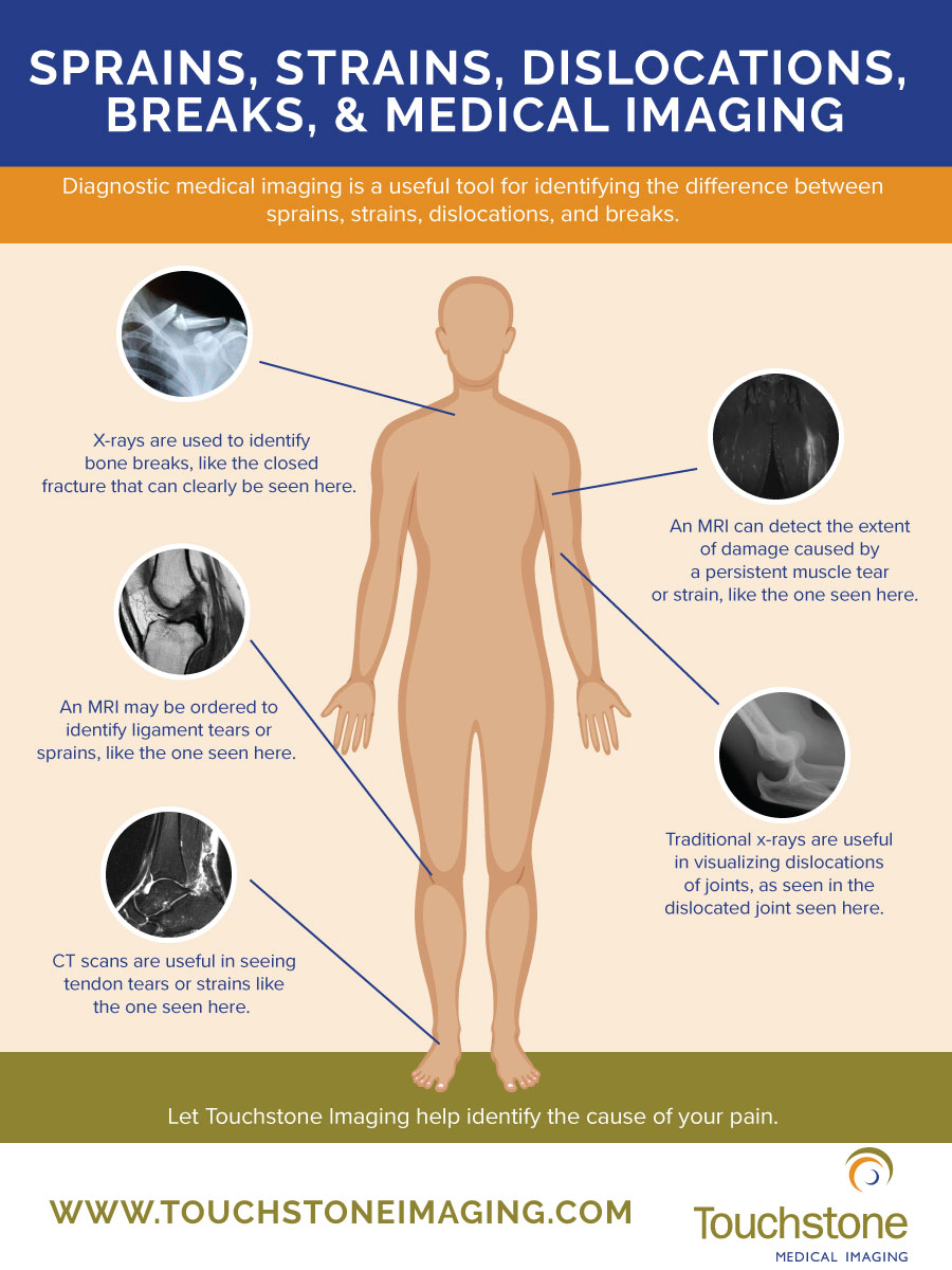
To accurately identify the injury and determine whether or not it is a sprain, you will likely first have an x-ray done. An x-ray provides a clear image of the more dense tissues on your body such as bone and thick ligaments. An x-ray will help identify or rule out any bone breaks or dislocations and may be able to show large ligament tears or detachments. If your injury cannot accurately be identified or a ligament needs to be further explored, you may be referred for a CT scan. A CT scan, or computed tomography, is a series of longitudinal x-rays that create a 3D image of your internal structures. A CT will allow your radiologist to clearly see a ligament tear and identify other soft-tissue injuries, much smaller than a traditional x-ray can reveal. Some providers may refer you for an MRI to identify very small tears or other structural anomalies.
Identifying Strains
A strain is an injury to either a muscle or to the tendon – the fibrous tissue that attaches muscle to bone. Strains are much less serious than sprains, but can cause similar pain and limited range of motion. Strains often heal much quicker than sprains and require less intervention. Just like with sprains, you can strain nearly any muscle or joint in your body and can injure multiple muscles or ligaments in the same area. The initial treatment is rest, ice, compression, and elevation along with some anti-inflammatory pain relievers. There is usually not much medical intervention required for recovery, but some people benefit from physical therapy. It is important to give your body time to rest and recover to prevent doing further damage or reinjuring the site.
Most times, medical imaging is not necessary for the diagnosis and treatment of a strain because the pain will be localized to the muscle affected and your provider should be able to determine a strain by the movements you are able or unable to make. However, for pain that persists or doesn’t improve, your provider may order a CT scan or an MRI to take a closer look at the soft tissues and evaluate micro-tears and the extent of the damage. For areas that have scar tissue build-up or have been damaged beyond what rest can repair, you may require surgical intervention.

Identifying Dislocations
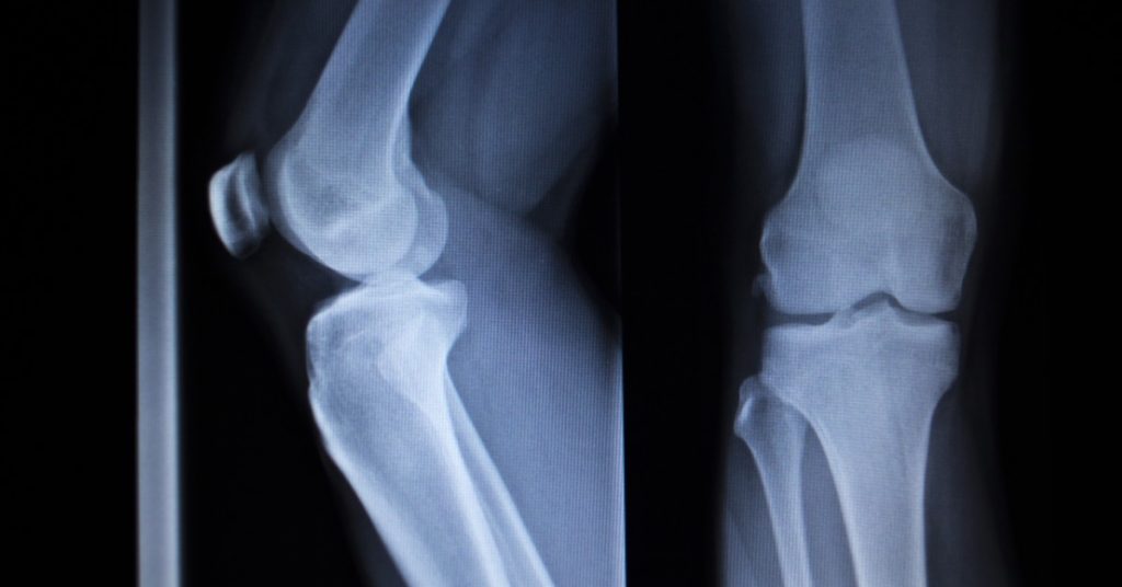
A joint dislocation is when a bone slips or is forced out of the joint. You can dislocate any joint in your body. When a joint dislocates, it is fairly easy to tell without medical imaging. The joint will be disfigured, you will experience excruciating pain, and there will be significant limitations in mobility and range of motion. Prior to reducing the joint, an x-ray may be obtained to ensure there are no additional injuries including tendon or ligament detachments or bone head breaks that will cause more damage when the joint is reset. A CT scan may be ordered to visualize nerve and blood vessel damage. An MRI may also be ordered, but is typically reserved for once the joint is back in place. Prior to the joint being reduced, it is important to keep it immobilized in place to prevent tendon, ligament, nerve, blood vessel, and muscle damage.
Identifying Breaks
A bone break is any compromise in the integrity of the bone and can range in severity from a stress fracture or hairline fracture to a complete break of the bone. In one of our previous posts, we dive into detail regarding the different kinds of bone breaks and how they appear on diagnostic imaging tests. To read more details about bone breaks, read our post here. When a bone breaks, symptoms may range in severity from pain and swelling, to complete loss of function in the area. You can break any bone in your body and there may be different kinds of breaks in the same bone or neighboring bones caused by a single injury. Initial treatment is to immobilize the injury as it lies to prevent further damaging the bone and surrounding muscle, nerves, and blood vessels.
A traditional x-ray is considered the gold standard in identifying bone breaks because the dense tissue of the bone readily shows on an x-ray. However, for hairline or stress fractures, an MRI may be done to assess the true extent of the damage and swelling. A bone scan may also be completed to get a better idea of the integrity of the bones. Your medical provider will use medical imaging results to safely reset the bone to allow for healing while minimizing additional damage.
If you have been injured and sustained a musculoskeletal injury, chances are your medical provider will order some form of diagnostic imaging procedure to evaluate the damage and develop a treatment plan. At Touchstone Imaging Centers, our locations offer x-ray, CT scans, MRI, and bone scans all taken by skilled technologists and interpreted by experienced radiologists. Our team of dedicated medical imaging professionals helps your team of treatment providers by providing them with quality images and results so they can provide you with the best treatment and hope for recovery. For all of your medical imaging needs, visit us online to find a Touchstone Imaging center near you.