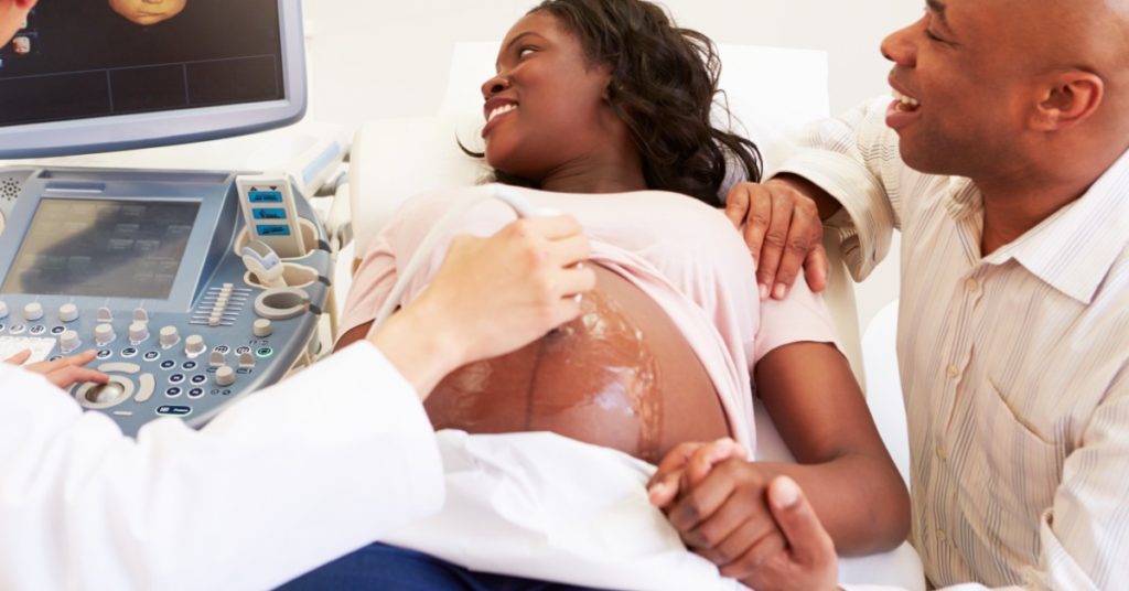
During pregnancy, you can count on at least two ultrasounds, the first one is to confirm pregnancy and the second, at around 20 weeks gestation, to assess the baby’s anatomy for normal growth and development. If it is a healthy pregnancy and baby, these should be the only two ultrasounds you receive during the duration of the pregnancy. Ultrasounds are the most common, and safest way to assess the things going on inside you and the status of the baby. Because ultrasound uses sound waves instead of radiation, it is accepted as a safe form of assessment for both mom and baby. If there are circumstances that require repeat or follow-up ultrasounds, you can bet it is in your best interest. Join us in today’s post as we discuss some of the various types of ultrasounds you may encounter during your pregnancy and reasons why your obstetrician may refer you for a repeat or follow-up ultrasound.
At Touchstone Imaging Centers, we provide ultrasound services at most of our locations including:
- Transvaginal ultrasound
- Traditional 2-D ultrasound
- 3-D Ultrasound
To learn more about these different ultrasounds, view our previous blog.
Routine Pregnancy Ultrasounds
During the first trimester, or when you make your first appointment, you should have a quick ultrasound that confirms pregnancy, looks for a heartbeat, assesses the number and location of sacs, and measures for approximate gestational age. Most providers prefer to wait until you are between eight and 12 weeks pregnant to get the best picture. This ultrasound can confirm multiple gestation and molar or ectopic pregnancies and will give you and your provider a good idea of what to expect in the coming months.
During the second trimester, between 20 and 23 weeks, you will have an anatomy scan. This ultrasound usually lasts much longer — about an hour — and will measure and assess all of the growing baby’s anatomy. At this point, the baby will have all of its organs, limbs, and digits, and the features are distinguished enough that malformations are readily evident. You’ll catch a glimpse of your baby’s facial features and be able to identify any structural abnormalities. Identifying issues or concerns at this point allows you and your obstetrician to formulate birth plans and discuss any treatments or ongoing testing moving forward. This scan will verify the estimated due date and status of the baby’s growth. This scan is also particularly useful in confirming the status of multiple babies and their wellbeing. This is also the ultrasound where you might be able to find out the sex of your baby.
Read more about the anatomy scan and what your obstetrician is assessing for here.
Common Reasons For Repeat Ultrasounds
If there is an abnormality noted on the anatomy scan or your baby’s position wouldn’t allow for complete visualization of all vital organs, you may have to repeat just the portion of the anatomy scan that assesses the organ in question. Once an abnormality is confirmed, additional repeat ultrasounds may take place in the third trimester, once fetal development is nearing completion. There are a great number of fetal abnormalities that a very common and may be corrected on their own as intrauterine growth and development takes place. It is important to get all the information and discuss result implications with your obstetrician.
Follow-up on abnormalities.
If there was a congenital abnormality identified on the anatomy scan, your obstetrician may want to reevaluate the situation later in pregnancy. Or, if the mother has a medical or gestational condition that may affect the development of the baby, third-trimester ultrasounds may be indicated. This will give a better idea of what to expect at delivery and guide resources you and the baby will need. Some common congenital abnormalities that may require reassessment as the pregnancy continues include (but are not limited to):
- Gestational hypertension in mother
- Gestational diabetes in mother
- Small or large for gestational age — monitor growth or reevaluate estimated due date
- Trisomy abnormalities
- Echogenic intracardiac foci
- Single umbilical artery
- Choroid plexus cyst
Identify hydramnios or oligohydramnios.
Excessive or reduced levels of amniotic fluid, respectively, can cause risks to both the mother and the baby. Your provider may want to assess fluid levels to determine fetal safety and dictate the need for modifying birth plans.
Identify the placental location.
A low lying placenta on an anatomy scan may appear to cover the cervix. During the final stages of pregnancy, this can cause a big concern of the exit is blocked for the baby. However, as the uterus expands to accommodate fetal growth, the placenta may migrate to a safe place. If your placenta still covers the cervix in the third trimester, you will be diagnosed with placenta previa, and a plan for c-section may be discussed.
Confirm intrauterine death.
If fetal demise is suspected, an ultrasound can be used to assess the absence of a heartbeat. Once fetal death is confirmed, you and your obstetrician will develop a plan for delivery.
Observe the fetal presentation.
While growing and developing, your baby may present in different ways and continue to move about. If your baby is breech — with the feet or buttocks closest to the cervix — at the second-trimester scan, they still have plenty of time to flip before it becomes a concern. As potential delivery nears, your obstetrician may order a repeat ultrasound to assess the presentation and then make plans for manually rotating the baby to a head-down position or discuss alternative birth methods.
Identify uterine and pelvic abnormalities of the mother.
At the second-trimester ultrasound, a few of the assessment checks include the mother’s anatomy. If there are uterine or pelvic abnormalities, uterine abnormalities, or an incompetent cervix, these things may affect the safe delivery of the baby and may need to be reassessed closer to delivery so appropriate birth plans can be discussed.
Other reasons not identified on previous scans.
Sometimes, events or symptoms in the pregnancy may warrant a follow-up ultrasound, even when things looked normal on initial scans. Some of these indications include:
- Pelvic or abdominal pain in the mother
- Lack of fetal movement
- Vaginal bleeding
- Abdominal trauma to the mother
- Infection during pregnancy
- Lack of maternal weight gain without explanation
- Poor advancement of uterine or abdominal growth in the mother
- Development of gestational diabetes
- Preeclampsia
- Medical emergencies
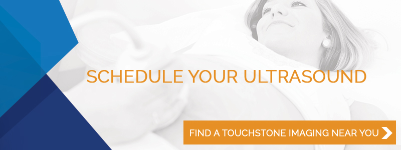
At Touchstone Imaging Centers, we know that you have a choice in where you have your medical imaging services performed. Our caring and experienced radiology technicians and radiologists work hard to provide the best patient experience and provide the most accurate results to your obstetrician. For all of your pregnancy-related ultrasound needs, find a Touchstone location near you and schedule your appointment today.
For more information, visit these other helpful Touchstone Imaging links: