
One thing that every new expecting parent waits impatiently for is the ultrasound images that allow a sneak-peek at their baking bundle of joy. The sonogram images offer a snapshot, and proof, that there is a baby and is one of the few ways that parents and their healthcare team can ensure that everything is going well. Although the images are something that families look forward to sharing with their loved ones and hanging on their refrigerators, they do serve a purpose. Although there is no set number of prenatal ultrasounds that will be performed during your pregnancy, here we will offer some insight into when baby scans are typically done and why.

Different Kinds of Prenatal Ultrasounds
When you get a prenatal ultrasound, there are a few techniques that may be used, so let’s familiarize you with them so you know what to expect.
A transvaginal ultrasound is performed by inserting an ultrasound wand (probe transducer) in the vagina to collect sonogram images. Transvaginal ultrasounds are used in early pregnancy when transabdominal scans would not yet be effective.
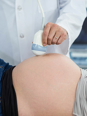
The traditional ultrasound is the standard transabdominal 2-D ultrasound that is most common. This type of ultrasound is used to create sonogram images of the developing baby and to capture a heartbeat.
3-D ultrasounds use special technology to generate 3-D images of the fetus. 3-D ultrasounds are not typically done in a medical setting unless there is a suspected problem but are offered by companies wanting to provide keepsakes for families.
4-D ultrasound is a step up from 3-D and can offer a peek at the features and movements of the baby. 4-D or dynamic 3-D ultrasounds are typically performed for novelty by expecting parents rather than for medical diagnostic purposes.
Fetal echocardiogram uses ultrasound waves to specifically assess the baby’s heart and blood flow in the event there are suspected congenital heart defects.
Confirmation of pregnancy.
Regardless of the suspected gestational age of the fetus, the purpose of the first ultrasound is to confirm pregnancy and the presence of a viable fetus. This ultrasound will confirm heartbeat, which depending on gestational age may not have an audible sound. The ultrasound technician will also measure the fetus to determine gestational age. Each woman will have the first ultrasound conducted at different times. Some obstetricians calculate gestation by the mother’s reported last menstrual period and will wait until the baby is approximately eight to 12 weeks old so that more medically meaningful images can be taken. During this scan, if there is no detectable heartbeat, don’t be concerned just yet, gestational aging by last menstrual period is not always accurate and a follow-up ultrasound will reveal more information. Additionally, it is about this time that the risk of spontaneous abortion is reduced by nearly 50 percent. In this first ultrasound, the fetus may appear to look like a gummy bear or a very small baby but is not likely to have distinguishable features aside from a head and possibly limbs.
In special circumstances, ultrasounds may be performed sooner or more frequently. For instance, with in-vitro fertilization, many reproductive specialists will order an ultrasound every week for a few weeks beginning between week five and eight. The purpose of these frequent ultrasounds is to ensure that the embryos implanted successfully in the uterine wall and that they are growing appropriately. High-risk pregnancies such as advanced maternal age or a previous history of abnormal pregnancy or loss of pregnancy may warrant more frequent and earlier baby scans. If a woman has abnormal vaginal bleeding or intense abdominal pain, an ultrasound may be performed to rule our molar or ectopic pregnancies, placental abruption, or fetal demise.
Nuchal translucency test.
Between weeks 11 and 13, pregnant women are offered an optional nuchal translucency (NT) test, which evaluates the probability of having a baby born with Down syndrome, trisomy 18, or congenital heart defects. The ultrasound is performed to measure the thickness of the back of the baby’s neck and can detect facial and limb deformities. Growth measurements can also be compared to the initial ultrasound to determine the rate of growth. This ultrasound, if conducted, will be accompanied by a blood test that will measure hormone and protein levels. This baby scan is completed transabdominally.
Anatomy scan.
Somewhere around week 20, you will be scheduled for an anatomy scan. This baby scan is conducted through the abdomen and tends to last longer than the other scans. During this ultrasound, the ultrasound technician will measure most of your baby’s body parts including the brain, head, heart, kidneys, limbs, and trunk. They will count the number of, now clearly visible, fingers and toes and look for facial abnormalities such as a cleft lip or palate. During this scan, the umbilical cord and amniotic fluid will also be visualized. Based on these baby scan results, your estimated due date may change because your obstetrician is able to compare the measurements to your initial scan and baby’s size will be much more accurate now.
Typically, the anatomy scan is the last scheduled ultrasound during pregnancy. However, in some cases, you may also receive a third-trimester scan. This is common in mothers of advanced maternal age, mothers who have gestational diabetes, low amniotic fluid, or pregnancy-induced hypertension (PIH), or whose baby measured either large or small during the anatomy scan. These follow-up scans are usually to monitor baby’s growth and development to ensure a safe pregnancy and delivery for both mother and baby. If there were abnormalities on the anatomy scan, a repeat scan may be scheduled for after week 30, as many congenital findings resolve naturally as the fetus continues to grow and develop. If you go beyond your due date, your obstetrician may order another ultrasound to make sure baby is doing well.
If you opt for additional ultrasounds, such as a 3D or 4D, those result in the best images between 25 and 30 weeks when the baby is small enough to capture a good portion in a single image and there is enough amniotic fluid for the ultrasound waves to travel through, capturing the best image. It is also at this time that baby’s features are fully developed and from this point on in the pregnancy baby will only change in appearance by gaining weight.
If your doctor orders a prenatal ultrasound for you or you are interested in getting a 3-D or 4-D ultrasound, consider Touchstone Imaging for your ultrasound needs. Our private medical imaging centers are small enough to ensure that you get the time and attention you deserve as opposed to the fast-paced, high patient turnover in a hospital radiology department. Yet, we are large enough to offer the latest technology and serve all of your medical imaging needs. We boast more than 25 years of radiology experience serving the residents of Texas, Colorado, Oklahoma, Nebraska, Florida and Arkansas.. Visit us online to find the nearest location to you and schedule your appointment!