Medical imaging is a powerful diagnostic tool that is used in conjunction with other diagnostic tests and procedures to confirm a diagnosis, develop an effective treatment plan, and follow-up to ensure the treatments worked and monitor progression or regression of chronic disease. The most common areas of the body that are imaged to reveal conditions or diseases are the chest, abdomen, and head. In today’s post, we are going to highlight chest scans and what they can reveal to an investigating medical team.
If you have visited your medical provider and been prescribed a medical imaging test to help find the cause of your concerns, trust the expert radiology team at Touchstone Imaging.
A Quick Anatomy Lesson
Before we jump right into the different medical imaging procedures that can be used to look inside the chest and what conditions they reveal, let’s take a quick minute to review some anatomy first.
The thoracic cavity is the space in your chest that is protected by your rib cage. Your ribs connect your spine to your sternum and give your body its shape and stability. More importantly, your thoracic cavity is home to many of your vital organs. Some of these structures include your esophagus, trachea, heart, lungs, diaphragm, thymus gland, aorta, spine, and a large portion or nerves, veins, and arteries.
Understanding just how much the chest holds may help make more sense of the number of things that can go wrong and how important medical imaging is to detect abnormalities. In a previous post, we discussed some of the things that a trauma surgeon is looking for when they order a chest CT, but this is just the tip of the iceberg!
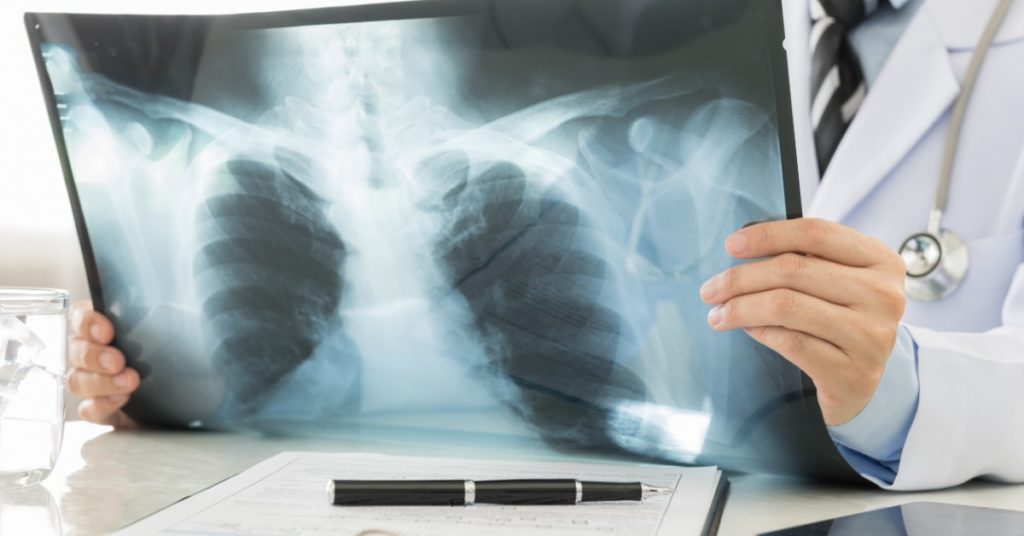
Chest X-Ray
Chest x-rays are usually conducted from a few different angles to capture a clear picture of the entire chest cavity. Some of the abnormalities or diseases that a chest x-ray can reveal include:
- Broken bones — collarbone, ribs, sternum
- Pneumonia
- Tuberculosis (TB)
- Lung tumor
- Cystic fibrosis
- Chronic obstructive pulmonary disease (COPD)
- Collapsed lung (atelectasis)
- Air (pneumothorax), blood (hemothorax), or fluid (pleural effusion in the pleural space (the space between the lung and the lung lining)
- Location of foreign objects
- Pacemaker, central line, port, intubation tube, and feeding tube placement
Computed tomography (CT)
A CT scan is a series of x-rays that can provide a clearer, multi-dimensional, multi-angle view of body structures. CT scans are typically used to take a closer look at abnormalities that are found on a traditional x-ray or to evaluate the extent of an obvious injury. Chest CT is effective at showing everything an x-ray can, plus:
- Tumors and nodules
- Bleeds
- Congenital abnormalities
- Lung disease
- Pulmonary embolism
CT angiography
CT angiography is a CT that uses a contrast dye to create images of blood vessels in the chest. This is done to look for pulmonary embolism and other narrowing or blockages in the blood vessels that go to the heart, lungs, brain, kidney, and limbs.
Mammogram
Mammograms and other women’s imaging that focuses on the chest can be used to reveal breast tumors. For more information on breast-specific diagnostic imaging, read our post that goes into depth about each procedure.
Chest MRI
An MRI of the chest is able to look at all of the structures in the chest including the soft tissues. Generally, a chest MRI is not the first diagnostic procedure that is done, but instead, is used to follow up on other diagnostic tests with questionable or less-than-clear results. Chest MRI can be used to:
- Detect size, and contents of masses
- View extent and spread of disease
- Grade cancer
- Assess the anatomy and function of the heart and its structures
- Assess lymph nodes
- Assess chest muscle and fat
- Look inside the bones
- Characterize lesions or masses identified on x-ray or CT
When your medical provider orders a medical imaging procedure of your chest, it is often done after a physical assessment, and in conjunction with other diagnostic procedures, including labs. The results of the imaging will be interpreted by a radiologist, and then your medical provider will put all the evidence together to form a diagnosis. It is important that you understand that a simple image of the chest is not enough to make a diagnosis and formulate a plan. This is why you do not have to have a doctor’s order to have a medical imaging test performed at Touchstone Imaging, but you do have to have a physician we can send the results to. Medical imaging is a powerful diagnostic tool, but is just a visual tool in the bigger picture of your health.
At Touchstone Imaging, our talented team of radiology specialists understands that your health matters and that the images we take make a difference. We ensure that every one of our images is high-quality and will result in the best interpretation. We are accredited by the American College of Radiology and recognized for MRI, CT, and mammography, as well as designated as a lung cancer screening center. When you need a diagnostic imaging test performed on your chest, visit us online to find a location near you and contact us to schedule an appointment!