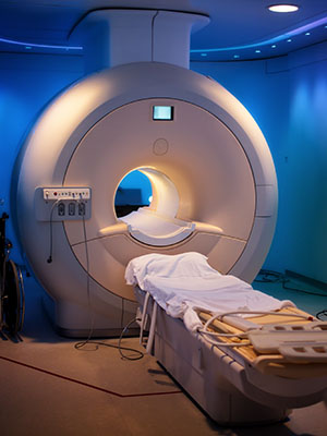


When your healthcare provider tells you they recommend a prostate MRI scan to help detect potential cancer early, you might be a little worried, but

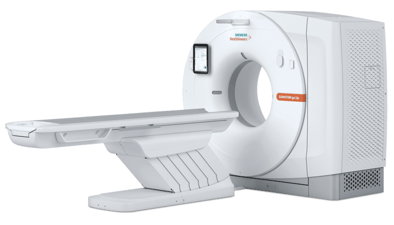
Being told you need a low-dose lung CT scan can leave you with questions, especially if you’ve never had this type of scan before. Whether

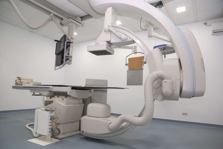
Learn more about why your healthcare provider has recommended a CT angiogram, what the scan can help detect, what to expect, and how to be prepared.