
When your healthcare provider examines your thyroid, and finds something concerning, one of the diagnostic tests they’ll order is a thyroid ultrasound.
The thyroid gland, located in your neck, plays a vital role in regulating your metabolism, growth, development, and other important functions.
An ultrasound will give your provider detailed images of your thyroid, which they will use to inform your care, based on your ultrasound results.
Let’s see exactly what you can expect from a thyroid ultrasound.
Why is a thyroid ultrasound important for detecting cancer?
A thyroid ultrasound is a non-invasive tool that uses sound waves to create images of your thyroid gland.
Ultrasound can help identify suspicious areas, such as nodules or lumps, in the thyroid gland that may not be easily found during a physical examination.
A thyroid ultrasound is particularly valuable for distinguishing between solid tumors, which have a higher risk of being cancerous, and fluid-filled cysts, which are usually benign (non-cancerous).
How does a thyroid ultrasound help my provider to look for cancer?
During a thyroid ultrasound, your healthcare provider looks for several indicators that might suggest the presence of cancer.
These include the size, shape, and composition of any thyroid nodules. Nodules with irregular borders, a taller-than-wide shape, or microcalcifications (tiny calcium deposits) may be more concerning for cancer.
The radiologist will also assess the blood flow to the nodule since increased blood flow can sometimes indicate cancer.
This detailed imaging allows the radiologist to determine which nodules may need further evaluation, possibly through a biopsy, to rule out or confirm cancer.
What should I expect during my thyroid ultrasound?
The process is quick, painless, and typically takes less than 30 minutes.
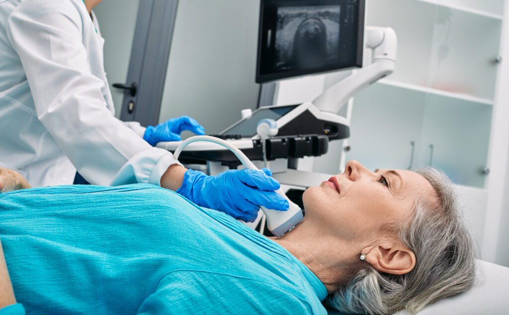
You’ll be asked to lie down with your neck extended to expose the thyroid area.
A small amount of gel is applied to your neck to help the ultrasound device, called a transducer, glide smoothly and ensure better contact with the skin.
A technologist moves the ultrasound device over your neck to capture images from different angles. You won’t feel any discomfort from the sound waves.
Afterwards, the technologist will wipe off the gel, and you can resume your normal activities immediately.
This non-intrusive approach makes thyroid ultrasound an ideal first step in evaluating thyroid health and investigating any concerns for cancer.
Identifying suspicious features in thyroid nodules
Your provider ordered a thyroid ultrasound because they want to take a closer look at your thyroid nodules.
As we’ll discover, thyroid nodules are an area of concern when it comes to diseases like cancer, and a thyroid ultrasound is great at providing clear images of them. We’ll help you understand why it’s so important for your provider to look for thyroid nodules.
Why are thyroid nodules a concern for cancer?
Thyroid nodules are small lumps that can form within your thyroid gland.
While most of these nodules are benign (meaning they are not cancerous), a small percentage can be malignant (which can be a sign of cancer).
Evaluating these nodules is crucial. Without detailed examination, it’s challenging to distinguish between nodules that are harmless and those that may pose a health risk.
Early detection through screening can lead to a better understanding of the nodule’s nature and, if necessary, more prompt treatment.
How will an ultrasound give my provider a better look at my thyroid nodules?
An ultrasound uses sound waves to create images of the inside of your body, offering a non-invasive look at your thyroid gland.
This allows a radiologist to see the size, shape, and number of nodules on your thyroid and report these findings to your healthcare provider for next steps in treatment or care.
Ultrasounds can provide detailed visuals that help in identifying characteristics of nodules that might be concerning.
For example, it can show if a nodule is solid or filled with fluid, which is an essential factor in assessing the potential for cancer.
This clarity is invaluable for helping your provider determine which nodules may need further evaluation.
How does ultrasound differentiate between benign and potentially cancerous nodules?
Ultrasound technology can accurately tell the difference between benign and potentially cancerous thyroid nodules.
When a radiologist looks at your ultrasound images, they’ll look for several different features that can indicate a higher risk of cancer, such as the presence of tiny calcium deposits.
Additionally, the ultrasound can assess the blood flow to the nodule; increased blood flow can sometimes be associated with cancer.
The radiologist will carefully analyze these features to decide whether a nodule is likely benign, or if it requires a biopsy for closer examination.
This step is crucial in the early detection and treatment of thyroid cancer, ensuring that any necessary action can be taken promptly and effectively.
Getting an ultrasound-guided thyroid biopsy
After an initial thyroid ultrasound, your healthcare provider may order an ultrasound-guided thyroid biopsy, especially if the ultrasound identifies areas of concern.
An ultrasound-guided thyroid biopsy is minimally invasive for you, it gives your provider actionable information, and it plays a crucial role in diagnosing thyroid conditions accurately.
Here, we’ll outline what you can expect from an ultrasound-guided biopsy of your thyroid gland.
Why would my provider recommend a thyroid biopsy?
A thyroid biopsy is typically recommended when your ultrasound results show nodules or irregularities that need a closer examination.
Not all thyroid nodules are cancerous, and in fact, the majority are benign.
However, the only way to conclusively determine the nature of these nodules is by taking a small tissue sample and analyzing it under a microscope.
This step is vital for your healthcare provider to make informed decisions about your health, ensuring any treatment plan is appropriately tailored to your needs.
What role does ultrasound play during a thyroid biopsy?
Ultrasound technology is not just for initial screenings. It’s also pivotal during a biopsy.
The ultrasound provides real-time images of your thyroid gland, guiding the physician in precisely locating the nodule of interest.
This live imaging is crucial for directing the biopsy needle to the exact location, minimizing discomfort and enhancing the accuracy of the tissue sample.
By using ultrasound guidance, providers can avoid areas that should not be disturbed, ensuring a safer and more effective biopsy.
How do biopsy results, combined with ultrasound, help to diagnose thyroid cancer?
Once the biopsy is complete and the tissue sample is analyzed, the results offer a clear picture of the cellular composition of the nodule.
These findings, when combined with the initial ultrasound imagery, provide a comprehensive understanding of the nodule’s characteristics.
Benign nodules have distinct features different from those potentially cancerous, and the combination of ultrasound imaging and biopsy results allows healthcare providers to diagnose with greater confidence.
If the biopsy reveals cancerous cells, your provider will have a detailed insight into the type and extent of the thyroid cancer, guiding the next steps in your treatment plan.
Monitoring, follow-up, and next steps
After your thyroid ultrasound, your provider will want to keep a close eye on your thyroid gland, especially if they observe thyroid nodules in your ultrasound results.
Your aftercare will likely involve further monitoring of your thyroid, which may include follow-up ultrasounds, depending on your results and your provider’s recommendations.
In this section, we’ll look at the importance of ongoing monitoring, and we’ll outline what your care plan may entail.
How is ultrasound used to monitor thyroid nodules over time?
Ultrasound can be used as a non-invasive and highly effective tool for monitoring thyroid nodules over time.
By providing clear images of the thyroid gland and its nodules, ultrasound allows your healthcare provider to track any changes in size, shape, or appearance of these nodules.
Regular ultrasound studies are key to observing the behavior of thyroid nodules, as some may grow or shrink over time, while others remain unchanged.
This ongoing monitoring is essential for determining the stability of the nodules or identifying any signs that may warrant further investigation or intervention.
Why is it important for my provider to monitor my thyroid after detecting nodules?
Monitoring your thyroid after the detection of nodules will help your provider to keep a close eye on any changes that may happen in the thyroid nodules.
This careful vigilance can significantly impact the effectiveness of treatment options, since it helps your provider find anything suspicious early.
Regular monitoring also provides peace of mind, knowing that any changes in your thyroid health are being meticulously observed and managed by your healthcare provider.
What are the next steps if an ultrasound or biopsy results suggest cancer?
Your healthcare provider will discuss the next steps, which typically involve a more comprehensive evaluation, and carefully putting in place a treatment plan to support your health and wellbeing.
Your next steps may include additional diagnostic tests to understand the extent of the cancer and to identify specific characteristics that could influence treatment choices.
Based on these findings, your provider will develop a personalized treatment plan, which may include surgery, radioactive iodine therapy, thyroid hormone therapy, or a combination of these treatments.
Your provider’s goal is always to choose the most effective––and least invasive––approach to managing and caring for your health.
How to schedule your ultrasound appointment with us
Touchstone Medical Imaging offers ultrasound scans in Arkansas, Colorado, Florida, Oklahoma, and Texas.
Reach out to us at Touchstone, and we’ll help you schedule a mammogram appointment at an imaging center near you, today.
We’re here to help you get the answers you need.

Frequently Asked Questions (FAQ)
It provides detailed images to help identify potential cancerous growths in the thyroid gland.
Expect a non-invasive study where a technologist glides a handheld device over your neck to capture images of your thyroid.
Because some nodules can be cancerous, but if they’re detected early, the condition can be treated.
It identifies characteristics like size, shape, and composition, which can indicate a higher risk of cancer.
If ultrasound findings suggest a nodule could be cancerous, a biopsy is needed for definitive diagnosis.
Ultrasound guides the needle precisely to the targeted nodule for a biopsy, enhancing accuracy and safety.
Regular ultrasounds track changes in nodules’ size and appearance, helping to assess risk or stability.
The next steps often include further evaluation, possibly another biopsy or surgery, guided by the specific findings and risk assessment.