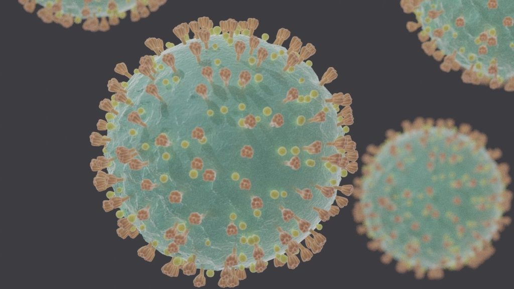
The outbreak of the COVID-19 Coronavirus has reached pandemic levels in a matter of mere weeks, taking thousands of lives and infecting thousands more around the world every day. In a valiant attempt to contain the spread of the virus and diagnose its pathology, medical imaging specialists are using CT scans and X-rays to study the lungs of infected patients.
The findings found so far are insightful and innovative, paving the way for what hopefully will soon become a vaccine for the novel coronavirus.
If you or someone you know is exhibiting symptoms of the virus, do your best not to panic. There is no need to be scared or concerned. A vaccine is coming soon, but in the meantime, we can do our part by following CDC guidelines and tips for staying healthy.
Note: Patients, your care and safety are our top priorities! We encourage you to follow CDC guidelines to self-quarantine if you’ve traveled to one of the high-risk countries or have been on a cruise in the last 14 days OR if you are experiencing fever, cough or shortness of breath.
CT Scan Findings
CT scans of COVID-19 patients have already demonstrated certain pulmonary characteristics indicative of the virus. Radiologists discerned clumps and patches of white in the lungs that they say resemble the pulverized glass of a window. As such, they refer to these patches as ground-glass opacities (GGOs).
The opacities are indicative of pneumonia, which seems to be the primary culprit behind the fatalities from coronavirus. Where air should normally be inhaled into the lungs, the CT scans show that patients of COVID-19 harbor a sticky mucus instead. This viscous substance causes breathing difficulties for the afflicted, especially those older in age or with a history of lung disease.
Other features of coronavirus found in the CT scans of the lungs include a “crazy paving appearance”, better understood by its technical nomenclature: inter- and intralobular septal thickening. Both the GGOs and the paving appearance have been found in bilateral, peripheral, and basal loci.
In addition to ground-glass opacities and abnormal appearance, the lungs of COVID-19 patients are characterized by air space consolidation, bronchovascular thickening and lesions, and traction bronchiectasis. Each is explained briefly below:
Air space consolidation: Also referred to as air space opacification, this is the symptom of COVID-19 coronavirus in which the lung parenchyma is visually subordinated to a foreign substance in the lungs. This is typically caused by pneumonia or related pulmonary issues, though it could also indicate lobar lung collapse.
Bronchovascular Thickening: This broad term can refer to one or more of a number of conditions seen in medical imaging. Namely, peribronchovascular interstitial thickening, bronchial wall thickening, and peribronchovascular consolidation. This anatomical anomaly may be seen with the use of CT scans or X-rays.
Traction Bronchiectasis: This condition refers to the permanent dilation of bronchi and bronchioles in the lungs, due either to pulmonary fibrosis or distorted architecture of the lung parenchyma. In either case, the bronchus is pulled away from its organic position by the traction of surrounding parenchymal fibrosis.
Note: One of the most important things to remember during this time is that panic will not solve anything. The best way to protect yourself and your loved ones from outbreak is simply by staying informed, taking care of your health, and following guidelines as they are issued by the CDC and WHO.
Prognosis
In many early cases of COVID-19, patients were dying at a mortality rate of 3%, though that number has since been reduced to an average of about 2%. No vaccine is currently available, though the combinatory treatment of protease inhibitors and antiviral agents has proven promising in mitigation or amelioration of the disease. Potential vaccines, if undergoing testing, are still in the preliminary phases of evaluation.
Infection Precautions for Medical Imaging Professionals
Medical imaging professionals working in close quarters with the novel coronavirus, whether in examining living patients or performing post-mortem scans, should adhere to certain protocols and precautionary measures to protect themselves from viral contraction.
The patients being examined should be placed under droplet-type precautions. This means medical masks, gowns, gloves, and protective eyewear. If a patient is receiving general radiography or X-rays, it’s best to administer the imaging portably; that is, conduct the radiographic procedure in the patient’s room so as to avoid transporting them. If a patient-accommodating x-ray is not feasible, the aforementioned droplet-type precautions should be implemented during mobilization to the site of testing.
Final Words
The novel coronavirus, COVID-19, has already been diagnosed in 873,767 cases (as of April 1, 2020) across dozens of countries. It is of the utmost importance that medical imaging professionals stay up to date on the latest findings and innovations in research of disease treatment.
If you or someone you know is showing any signs of the coronavirus, please follow the guidelines provided by the CDC here. Please do not visit our clinic if you have been symptomatic or traveled outside of the country in the last three weeks.
For more information on CT scans, X-rays, and general diagnostic medical imaging, please reach out to Touchstone Medical Imaging today.