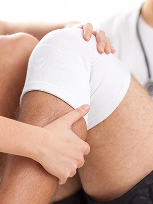
If you have joint pain or your doctor suspects a joint problem not seen on a regular MRI, he or she may order an arthrogram. You may be unfamiliar with this type of medical imaging and will want to know what to expect at your appointment. This column will explain what an arthrogram is, what it is used for, and what to expect during the exam.
Preparation
No special preparation is needed. You do not need to restrict food or water beforehand unless you are directed to do because of sedation. After marking the injection spot (with the aid of the fluoroscope) and cleansing the area with antiseptic, you will receive a local anesthetic to help deaden the small pain from the joint injection. After the anesthetic has taken effect, a longer needle will be inserted into the joint and is guided by fluoroscopy into the correct position. Following this, a fluid mixture containing special MRI or CT contrast will be injected. The needle will then be removed, and you will move the joint to distribute the dye throughout the space. The joint may feel full during this time, but there should be little pain.
MRI or CT Arthrography
After the contrast solution is injected into the joint, you will be taken to either the MRI (most likely) or the CT (if going into the MRI is contraindicated) room for scanning. This can be up to 30 minutes for an MRI or as little as 5 minutes for a CT. After the scan, a computer processes the images and the radiologist interprets them, sending a report to your provider. You usually will take a CD copy of the scan with you for your provider to look at if desired.
Who Needs Arthrography?
If you have ongoing joint pain that is not being relieved, swelling of a joint, or limited mobility, you may need further imaging. Usually, a regular MRI (which is usually all that is needed) is performed. If an answer is not found with the regular MRI, or if the orthopedic physician suspects certain types of joint tears or problems, he or she may order an arthrogram. Problems can be found in the joint capsule, ligaments or cartilage which may not be obvious on the regular MRI, including tears and cartilage degeneration.
After an Arthrogram
Because of the injection of the contrast dye, you may experience swelling in the joint area, and you may have aching pain or discomfort. Ice and anti-inflammatory painkillers can be used to alleviate any symptoms, which usually will disappear after 48 hours. There can be a rare risk of dislocation of the joint after your procedure, so vigorous exercise such as lifting heavy weights after an arthrogram should be limited. Once any pain is gone, you can resume normal activities. Contact your physician if your symptoms persist past two days, or if you have any concerns. You will receive your results from your provider, and you may be advised to follow up with medication, physical therapy, or surgery.
Arthrography is incredibly useful in diagnosing joint problems and finding a treatment plan that works. If you are experiencing limited range of motion in a limb, or are having continual shoulder or knee pain, you may be a good candidate for arthrography. When you need to have medical imaging done, know that you can choose where to go, and you don’t have to go to the hospital or its (usually) more expensive imaging center. Contact us to schedule an arthrogram at a Touchstone Imaging center near you.


