
A stroke is a medical emergency caused by the interruption of blood supply to the brain. The reduction of blood to a part of the brain deprives the tissues of much-needed oxygen and nutrients. Within just minutes, brain cells begin to die. When circulation is restored, some of the cells may recover, but there are usually some residual symptoms.
A stroke can be caused by either a blockage of blood flow to the brain or by free blood in the brain. Blockages of blood flow in the brain are caused by a clot or narrowing of blood vessels and are referred to as an ischemic stroke. Blood in the brain is usually caused by blood vessels in the brain breaking and leaking blood into the surrounding tissues, it is referred to as a hemorrhagic stroke. Nearly 87% of all strokes are ischemic and are a result of cardiovascular disease.
The brain is a vital organ that controls all of our thoughts and bodily functions. When part of the brain suffers cell death, it can create significant symptoms that manifest in various, but obvious ways. Left untreated (or delay treated), stokes can cause permanent damage or brain death. Nearly 800,000 Americans will suffer from a stroke this year, and nearly 75% of those victims will be over the age of 65. Stroke will be the cause of death for about 140,000 Americans and remains the number one cause of disability in the United States. Fortunately, due to the advancing technology of medical imaging and early medical intervention, more and more people are not only surviving strokes, but regaining function after a stroke when action is taken quickly.
When it comes to treating a stroke, time is of the essence and not a minute can be wasted. To help improve outcomes, quick access to high-quality medical imaging cannot be understated. Join us in today’s post as we review the anatomy of a stroke and how medical imaging can make the difference.
Medical Imaging That Guides Stroke Treatment
MRI and CT technology are both great options for visualizing the brain in great detail. However, because of the time it takes to complete, the CT scan remains the gold standard for acute stroke management. While MRI can take a better, in-depth look at the brain tissue, the procedure takes between 45 minutes and an hour, and is not great for detailing an active event. MRI is useful for post-intervention brain mapping to gain a baseline and detail the extent of damage to formulate a treatment plan and prevent recurrent strokes.
It is important to have quick access to medical imaging to identify not only what kind of stroke the patient is having, but where in the brain it is. As we mentioned, previously, time is of the essence when treating stroke and the treatments for ischemic and hemorrhagic stroke are dramatically different. However, since both types of stroke cause similar symptoms, it is impossible to tell which kind a patient is suffering without reliable medical imaging. If imaging is skipped and a stroke is treated blindly, the treatment for an ischemic stroke can worsen a hemorrhagic stroke and vice versa.
CT Scans Reign Supreme
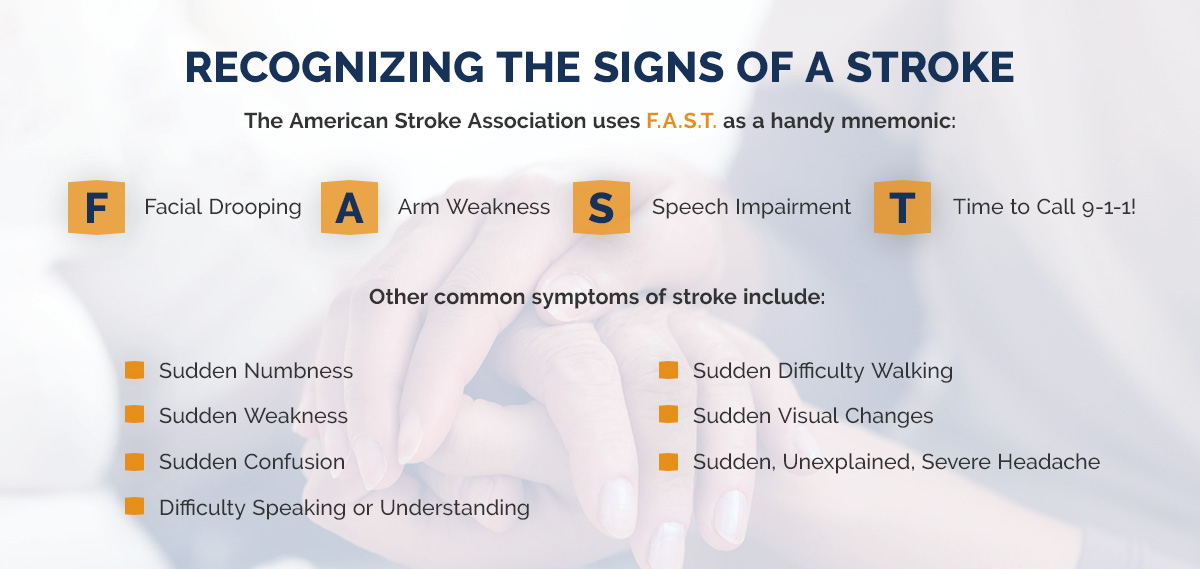
Noncontrast computed tomography is a quick diagnostic procedure that allows radiologists and medical providers to quickly visualize the brain and identify blocks or bleeds to allow the stroke medical team to perform quick interventions and minimize damage. CT scans are useful at identifying early stroke symptoms and damage, which is why it is important to never delay seeking medical treatment if you experience any of the symptoms listed in the image above. CT scans have proven to be useful in distinguishing between ischemic and hemorrhagic strokes or other conditions that may mimic strokes, including brain tumors. CT scans are useful in distinguishing between blood and normal cerebrospinal fluid, which is helpful during a hemorrhagic stroke.
 CTA Offers a Closer View

Computed tomography angiogram (CTA) is a CT scan that is performed with a rapid injection of contrast dye that allows CT imaging to isolate the blood vessels in the brain. This is particularly useful in discovering where the issue is and how severe it is. At the same time as a CTA, a CT perfusion (CTP) can be performed to identify blood flow and perfusion in the brain. Gaining a clear picture of blood flow in the brain during a stroke can help guide treatment and develop the best-individualized treatment plan for the patient. With a clear picture of the vessels that are involved, the medical treatment team can administer care more confidently.
For more information about post-stroke medical imaging intervention, check out these resources.
Imaging in Acute Stroke
Imaging of Ischemic Stroke
State-of-the-Art Imaging of Acute Stroke
At Touchstone Imaging Centers, our skilled imaging technologists are trained and proficient in performing all of the above mentioned medical imaging procedures. Because our imaging clinics are free-standing clinics, we are not often involved in acute stroke imaging and we would not encourage you to stop by our location near you if you are experiencing active stroke symptoms. However, we are more than happy to be involved in your on-going post-stroke management. For answers to all of your medical imaging questions or concerns, don’t hesitate to contact our team. To schedule your follow-up brain MRI or CT scan, contact a Touchstone Imaging center nearest you.