Medically speaking, being a woman is a difficult task. From a reproduction cycle that seems to take no mercy on the host to a variety of chronic diseases and conditions that disproportionately afflict women, it is a wonder how we have a longer lifespan than our male counterparts. [To validate the menfolk here, there are many chronic illnesses and conditions that afflict men greater than women and cancers in body parts that women lack that make being a man difficult as well.] Women’s bodies are built to carry and care for children and the hormones and nutrients that make this possible often wreak havoc on the woman’s own body and makes it more prone to autoimmune diseases and cancers. For this reason, it is important for women to keep up with routine screenings to ensure a long, healthy, comfortable life.
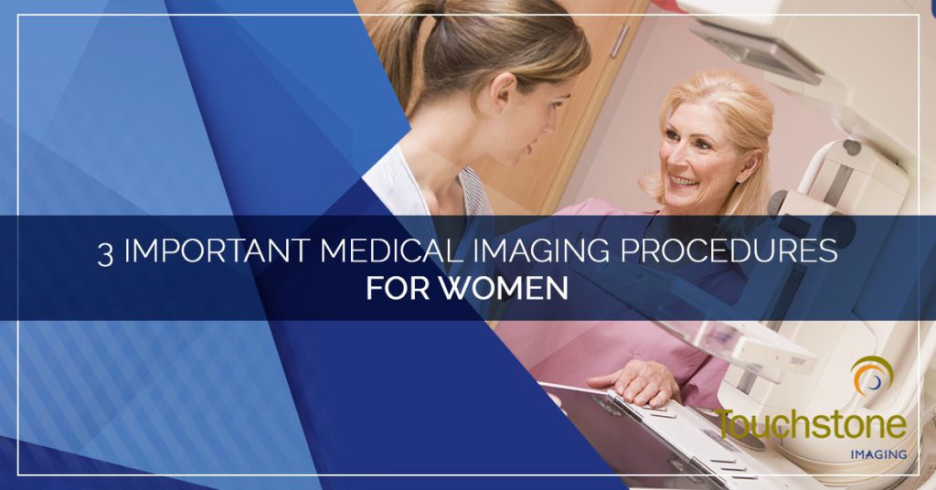
Due to the nature and prevalence of the conditions that affect women, specialty medical imaging services have been tailored to the specific needs of women. Follow along today as we discuss the top three medical imaging procedures that a majority of women will use at some point in their lives.

Ultrasound
Ultrasound is a useful non-invasive diagnostic imaging tool that does not use radiation or magnetic waves. Because it is safe to use on a fetus, it is the medical imaging tool of choice for pregnant women and to monitor a pregnancy. During pregnancy, an ultrasound will be used several times to monitor growth and ensure the proper development of the unborn baby.
Even if a woman never has a pregnancy, an ultrasound may be used to view internal reproductive organs including the cervix, ovaries, and uterus. An ultrasound of these organs can help detect masses or areas of concern that cannot otherwise be easily visualized.
 Breast Imaging
One in eight women will be diagnosed with breast cancer at some point in their life. While more than 80% of these women will survive to tell their grandchildren about their battle, that is in large part due to the ability of modern medical imaging modalities to detect and identify breast cancer earlier and more reliably. Because each breast is as unique as the woman it is attached to, there have been significant advances in the world of medical imaging to accommodate every breast.
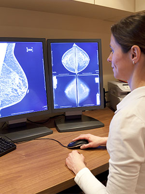
Screening Mammogram
A screening mammogram is the standard wellness exam that is covered under most insurance plans as preventative. A series of x-rays are used to produce images of the breast tissue to identify areas of greater density — tumors. At Touchstone Imaging, you don’t even need a physician referral to have a screening mammogram performed (however, since we are not a treatment center, we do require a provider to send the results to).
Breast MRI
A breast MRI can be used as supplemental imaging in addition to a screening mammogram. A breast MRI can be used to detect and classify breast diseases, including cancer. This modality is useful for identifying ruptured breast implants, differentiating tissues, and staging cancer.
Breast Ultrasound
A breast ultrasound is another useful supplemental imaging tool that can be used in conjunction with a mammogram. Because it is difficult to get a complete picture all at once, the ultrasound should not replace the mammogram, but it can be used as an investigative tool. Ultrasounds are great for examining palpable lumps and dense breast tissue. For lumps that can be felt and easily seen on an x-ray, an ultrasound can help determine if the mass is fluid-filled — lymph tissue, infected mammary gland, or a cyst — or is a solid mass before a determination is made regarding whether a lumpectomy is necessary.
Breast Tomosynthesis
Breast tomosynthesis, more commonly referred to as 3D mammography, is the newest in breast cancer screening technology. A 3D mammogram combines multiple x-ray images to create a 3D image as opposed to the traditional 2D image. This allows much smaller masses to be identified much sooner than other modalities can offer. Because of the multiple angles captured simultaneously, the 3D mammogram can also better detect masses in dense breast tissue.
Bone Density Scan
Bone density scans are not exclusive to the female population, but because osteoporosis and other bone density issues affect women at a greater rate than men — 1 in 3 as compared to 1 in 5, respectively — the bone density scan technology is used for women more frequently, and most women will have a bone scan during their life. As we discussed earlier, hormone and vitamin changes and deficiencies are more common in women, especially in those who go through pregnancy and menopause. Bone density scans are used to evaluate bone loss and fragility to help get patients the care they need at an early stage and provide helpful treatment before a condition worsens.
At Touchstone Imaging, our skilled technicians understand the specialized imaging needs of women. We staff women at our women’s imaging centers to help put you at ease while you are getting Top Rated Local® care from an American College of Radiology (ACR) accredited imaging center. When you need women’s imaging services, visit us online to find a Touchstone Imaging location near you and schedule your appointment today.